Molecular Imaging

Molecular imaging (also called nuclear medicine or nuclear imaging) can image the function of cells inside the body at the molecular level. This includes the imaging modalities of positron emission computed tomography (PET) and single photon emission computed tomography (SPECT) imaging. How does PET and SPECT imaging work? Small amounts of radioactive material (radiopharmaceuticals) injected into a patient. These can use sugars or chemical traits to bond to specific cells. The radioactive material is taken up by cells that consume the sugars. The radiation emitted from inside the body is detected by photon detectors outside the body. Computers take the data to assemble images of the radiation emissions. Nuclear images may appear fuzzy or ghostly rather than the sharper resolution from MRI and CT.  But, it provides metabolic information at a cellular level, showing if there are defects in the function of the heart, areas of very high metabolic activity associated with cancer cells, or areas of inflammation, data not available from other modalities. These noninvasive imaging exams are used to diagnose cancer, heart disease, Alzheimer’s and Parkinson’s disease, bone disorders and other disorders. 
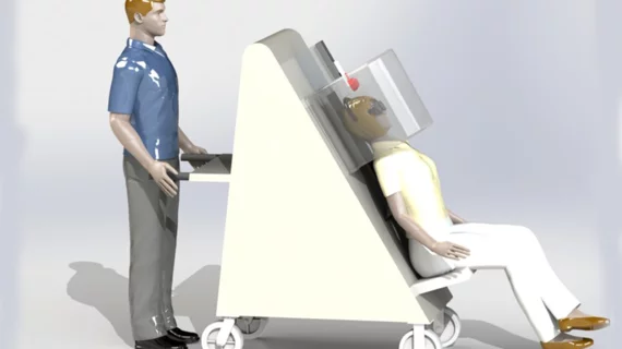
Frost And Sullivan applauds Gamma Medica for improving the quality and efficiency of breast cancer diagnosis with its unique LumaGEM MBI System

Based on its recent analysis of the breast imaging systems market, Frost and Sullivan recognizes Gamma Medica with the 2014 Global Frost and Sullivan Award for Product Leadership.

Thumbnail

Big data highlights new Parkinson’s variants

A handful of new genetic variants point to Parkinson’s disease, the National Institutes of Health (NIH) announced yesterday. The six previously unreported risk factors will contribute to researchers’ understanding as they work to develop better treatments for the neurodegenerative disease.

Thumbnail

Alzheimer’s therapies could result in $12B market by 2023

The industry for Alzheimer’s drug treatments is expected to grow from modest beginnings to big business in the next decade, according to a June report by Decision Resources Group.

Thumbnail

Photoacoustic PET takes imaging inside GI tract

A novel, non-invasive molecular imaging technique uses nanotechnology to allow clinicians a view into the gastrointestinal track without having to resort to invasive procedures, according to a study published July 6 in Nature Nanotechnology.

Radiocholine PET/CT has a niche in prostate imaging

Selection of patients with advanced prostate cancer who are eligible for salvage radiation therapy could be improved with the use of radiocholine PET/CT, according to a review of recent research published today in the Journal of Nuclear Medicine.

Not just three scans: Understanding FDG PET coverage

The Centers for Medicare & Medicaid Services provided a reiteration of the agency’s national coverage decision regarding oncologic FDG PET this summer and clarified specifically the –KX modifier that needs to be used when clinicians code for the fourth scan and beyond.

Thumbnail

Neuro study of worms is a 'brain hack'

Nematode worms are providing a very, very small window on the neural connections that we humans share. A feature in New Scientist follows the circuitous path of our knowledge about consciousness and the neural networks of the brain.

Plumbing the depths of PTSD

A feature in The New Yorker explores how Daniela Schiller, PhD, from the Mount Sinai School of Medicine’s Brain Imaging Core in New York City, has approached a kind of memory erasure for people who have suffered greatly from post-traumatic stress disorder (PTSD) with the aid of cutting-edge neuroimaging.

Around the web

Positron, a New York-based nuclear imaging company, will now provide Upbeat Cardiology Solutions with advanced PET/CT systems and services. 

The nuclear imaging isotope shortage of molybdenum-99 may be over now that the sidelined reactor is restarting. ASNC's president says PET and new SPECT technologies helped cardiac imaging labs better weather the storm.

CMS has more than doubled the CCTA payment rate from $175 to $357.13. The move, expected to have a significant impact on the utilization of cardiac CT, received immediate praise from imaging specialists.