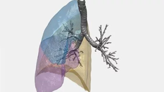
Computational method uses CT, algorithms to assess lungs for COPD
A research team compiled of mathematicians, clinicians and imaging technicians from the University of Southampton in the U.K. recently developed a computational method to analyze x-ray images of the lungs for overall function and the presence of fatal diseases.
According to a May 10 university press release, the multi-disciplinary team's method is rooted in topology, a branch of mathematics that studies complex shapes, and utilizes CT scans alongside high-performance computing and algorithms to better diagnose chronic obstructive pulmonary disease (COPD) and other lung diseases.
The study was published in Nature Scientific Reports on March 28.
“Our study shows that this new method, employing topological data analysis, can complement and expand on established techniques to give a valuable, accurate range of information about the lung function of individuals," said lead researcher Jacek Brodzki, PhD, a mathematics professor at the University of Southampton, in a prepared statement. "Further research is needed, but this could eventually aid decisions about the treatment of patients with serious, or potentially serious, lung conditions.”
The researchers computed numerical characteristics, in 3D, of the entire bronchial trees of 64 patients categorized as healthy non-smokers, healthy smokers, patients with moderate COPD or patients with mild COPD.
Brodzki and colleagues then analyzed the structure and size of the bronchial tree, the length and direction of its branches, and changes in shape when the patient fully inhaled and exhaled.
A larger, complex bronchial tree resulted in better overall function. In contrast, the smaller and more distorted the tree was, the poorer the lung function.
“This method is a major advance in our ability to study the structural abnormalities of COPD, a complex disease that affects so many people and, sadly, results in significant morbidity and mortality," said Ratko Djukanović, a professor of medicine at the University of Southampton and senior investigator National Institute for Health Research, in a prepared statement.

