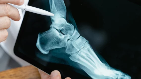
Orthopedic imaging relies on X-ray, MRI and CT to diagnose disorders and injuries affecting the bones, muscles, ligaments, tendons, cartilage, and spine. Orthopedists also use these test results to create an effective treatment plan.
Experts hope the information gained from their research could help providers better determine whether patients will regain mobility after sustaining an injury.
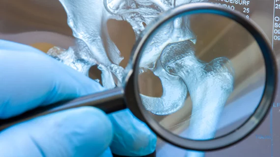
Many people with low bone mineral density are not aware of their condition because they fail to get screened through DXA or are not eligible due to age.
The technology is so promising that it is being integrated into GE Healthcare’s MRI scanners so that providers can conduct more thorough evaluations of how these implants hold up over time.
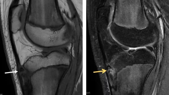
Although gadolinium-based contrast agents are largely considered safe and are routinely used for MRI exams, experts suggest that providers should still utilize GBCAs sparingly for musculoskeletal studies.
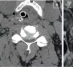
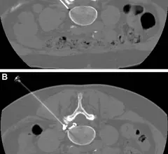
In PRF procedures, a probe is used to intermittently apply energy directly to the dorsal root ganglia, which is often where pain and neurologic symptoms associated with sciatica originate.
Although rare, SIRVA made its way into headlines following the widespread rollout of COVID vaccines. This prompted a renewed push among the medical community to better define the side effect.
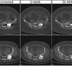
Such scans offer significant reductions in radiation exposure, but there is concern that lower dosage might sacrifice image quality, causing readers to miss important findings.
Clinicians have been using HeartSee to diagnose and treat coronary artery disease since the technology first debuted back in 2018. These latest updates, set to roll out to existing users, are designed to improve diagnostic performance and user access.
The cardiac technologies clinicians use for CVD evaluations have changed significantly in recent years, according to a new analysis of CMS data. While some modalities are on the rise, others are being utilized much less than ever before.