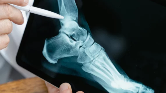
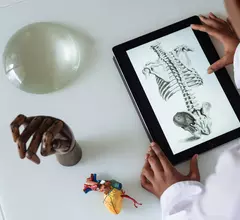
Orthopedic imaging relies on X-ray, MRI and CT to diagnose disorders and injuries affecting the bones, muscles, ligaments, tendons, cartilage, and spine. Orthopedists also use these test results to create an effective treatment plan.
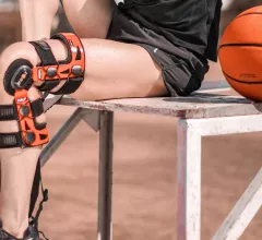
Experts hope the information gained from their research could help providers better determine whether patients will regain mobility after sustaining an injury.
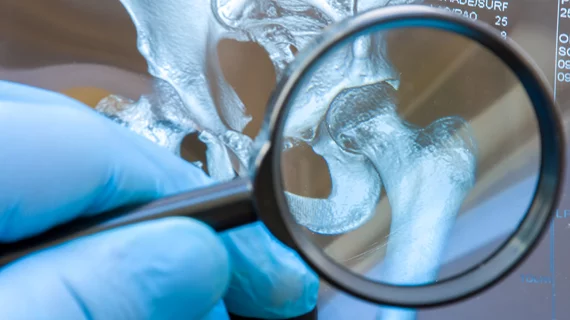
Many people with low bone mineral density are not aware of their condition because they fail to get screened through DXA or are not eligible due to age.
The technology is so promising that it is being integrated into GE Healthcare’s MRI scanners so that providers can conduct more thorough evaluations of how these implants hold up over time.
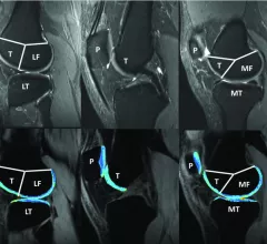
Many decision support tools catered to knee osteoarthritis have emerged in recent years, but external validation that ensures these algorithms can operate in a clinical setting has been lacking.
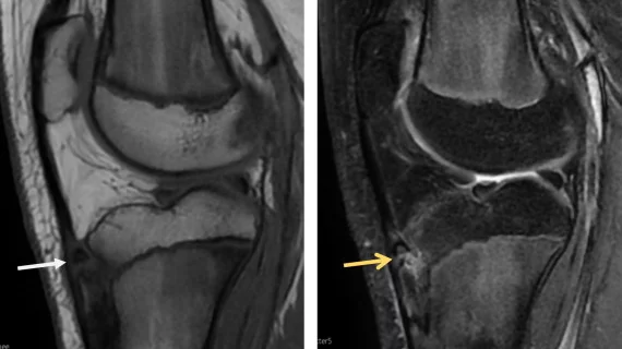
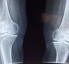
Reperforming lateral knee radiographs is common practice but consumes unnecessary resources and exposes patients to added radiation, experts explained in Radiography.
The University of Texas Medical Branch and College of Health Care Professions kicked off the program in January and both sides see a bright future ahead.
Clinicians have been using HeartSee to diagnose and treat coronary artery disease since the technology first debuted back in 2018. These latest updates, set to roll out to existing users, are designed to improve diagnostic performance and user access.
The cardiac technologies clinicians use for CVD evaluations have changed significantly in recent years, according to a new analysis of CMS data. While some modalities are on the rise, others are being utilized much less than ever before.