Orthopedic imaging relies on X-ray, MRI and CT to diagnose disorders and injuries affecting the bones, muscles, ligaments, tendons, cartilage, and spine. Orthopedists also use these test results to create an effective treatment plan.
Experts hope the information gained from their research could help providers better determine whether patients will regain mobility after sustaining an injury.
Many people with low bone mineral density are not aware of their condition because they fail to get screened through DXA or are not eligible due to age.
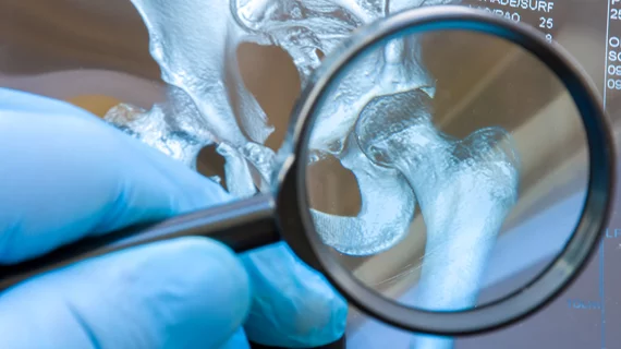
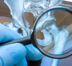
The technology is so promising that it is being integrated into GE Healthcare’s MRI scanners so that providers can conduct more thorough evaluations of how these implants hold up over time.
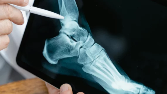
Established clinical guidelines hold that patients presenting with ankle issues should not receive advanced imaging ahead of standard radiography. New research shows a substantial proportion of ordering clinicians sending these patients straight to MRI anyway.
“These findings emphasize the importance of early recognition of IPV and timely intervention to prevent further harm to the victim,” authors of research published in Academic Radiology cautioned.
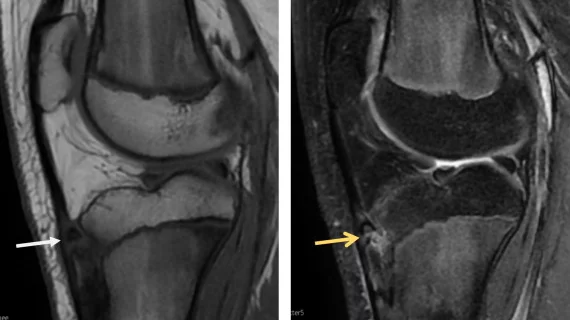
Because sacral insufficiency fractures do not always show a fracture line, they can be difficult to diagnose or even misdiagnosed as bone metastasis, which could result in additional treatments like radio-chemotherapy.
A study in Lancet Digital Health reports that a previously validated, high performing AI model committed troublesome errors when confronted with atypical anatomy.
Experts urged physicians to take extra protective measures when in the presence of metal protheses during procedures and to be vigilant in shielding their eyes from additional exposure.
Research published recently in Radiology found comparable sensitivity and specificity between artificial intelligence and clinicians for fracture detection.
Clinicians have been using HeartSee to diagnose and treat coronary artery disease since the technology first debuted back in 2018. These latest updates, set to roll out to existing users, are designed to improve diagnostic performance and user access.
The cardiac technologies clinicians use for CVD evaluations have changed significantly in recent years, according to a new analysis of CMS data. While some modalities are on the rise, others are being utilized much less than ever before.