Ultrasound

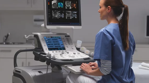
Ultrasound, also referred to as sonography or diagnostic ultrasound, uses high-frequency sound waves to visualize soft tissue. Ultrasounds are frequently ordered to measure fetal anatomy during pregnancy, check for blood clots and to guide needle biopsy procedures of the breast, abdomen and pelvis. The imaging modality does not use any radiation to create images. Find news specific to cardiac ultrasound (echocardiography).

Imaging suggests blood clots are more common in COVID than pneumonia

In a recent study, venous thromboembolisms were detected in those with COVID via multiple imaging modalities at a rate of 16.3% compared to 9.2% in individuals with CAP.

ovaries ovarian cancer

These ultrasound features predict ovarian cancer

Doctors created a simple system that categorizes adnexal lesions as classic or nonclassic to streamline the process of ovarian cancer risk stratification.

Research links echocardiographic measures to dementia risk

For the first time, associations between left atrial function and dementia were uncovered via electrocardiograms, experts shared in JAMA.

William A. Zoghbi, MD, MACC, FAHA, FASE, is the chair of the Department of Cardiology at the Houston Methodist DeBakey Heart and Vascular Center, and past president of both the American College of Cardiology (ACC) and the American Society of Echocardiography (ASE).

VIDEO: New advances in echocardiography

William A. Zoghbi, MD, past president of the ACC and ASE, discussed the latest trends in cardiac ultrasound technology. 

Thumbnail

Ultrasound features that indicate difficulty of vaginal childbirth

Ultrasound assessments of fetal head position have been used to predict delivery mode, but never delivery difficulty, experts recently discussed in the Journal of Gynecology Obstetrics and Human Reproduction.

Thumbnail

Specialized ultrasound can accurately detect prostate cancer, new research shows

Ultrasound pinpointed 4.3% fewer prostate cancers in comparison to multiparametric MRI, according to new research published in Lancet Oncology.

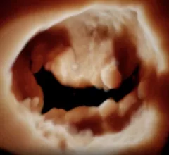
The mitral valve seen on 3D cardiac ultrasound offers a noninvasive surgeon's view of the valve for better diagnostics and pre-planning of structural heart procedures. Example from the GE Vivid E95 FlexiLight photo-realistic 3D/4D lighting system.

Q&A: William Zoghbi breaks down the future of echocardiography

In a new interview, former ACC and ASE President William A. Zoghbi, MD, reviews some of the biggest trends in cardiac ultrasound.

Thumbnail

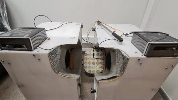
Ultrasound images reveal how smoking before conceiving impacts embryonic development

"The delay in morphological development cannot be fully recuperated during the course of the pregnancy, as is shown by the 20-week ultrasound scans and birth weights,” experts cautioned in Human Reproduction.

Around the web

Positron, a New York-based nuclear imaging company, will now provide Upbeat Cardiology Solutions with advanced PET/CT systems and services. 

The nuclear imaging isotope shortage of molybdenum-99 may be over now that the sidelined reactor is restarting. ASNC's president says PET and new SPECT technologies helped cardiac imaging labs better weather the storm.

CMS has more than doubled the CCTA payment rate from $175 to $357.13. The move, expected to have a significant impact on the utilization of cardiac CT, received immediate praise from imaging specialists.

Trimed Popup
Trimed Popup