Ultrasound

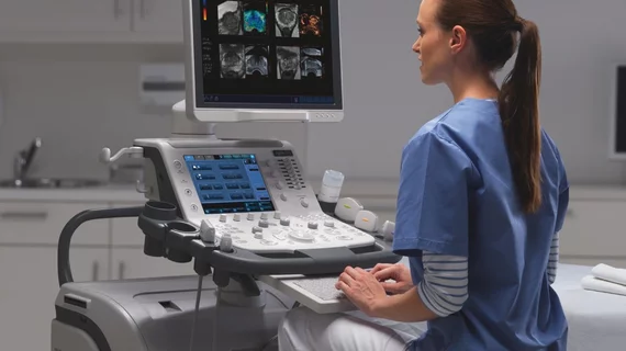
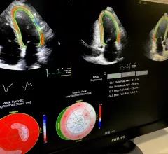
Ultrasound, also referred to as sonography or diagnostic ultrasound, uses high-frequency sound waves to visualize soft tissue. Ultrasounds are frequently ordered to measure fetal anatomy during pregnancy, check for blood clots and to guide needle biopsy procedures of the breast, abdomen and pelvis. The imaging modality does not use any radiation to create images. Find news specific to cardiac ultrasound (echocardiography).

Thumbnail

Prior cardiotoxicity linked to 30% increased risk of CHF during pregnancy

Women with a history of cardiotoxicity from previous cancer treatments are around 30 percent more likely to experience clinical congestive heart failure (CHF) before, during or after pregnancy, according to research published ahead of print in the Journal of the American College of Cardiology.

Thumbnail

Jonathan Lindner elected president of the American Society of Echocardiography

The 17,000-member American Society of Echocardiography (ASE) named Jonathan R. Lindner, MD, president of the organization during its 29th annual scientific sessions in Nashville, Tennessee.

Thumbnail

FDA-approved AI echocardiogram software bests cardiologists in reducing LVEF variability

A deep-learning software that can automatically calculate left ventricular ejection fraction (LVEF) with less variability than a cardiologist recently received approval from the U.S. Food and Drug Administration (FDA).

New registry seeks to improve cardiovascular ultrasounds

ImageGuideEcho—the first registry devoted solely to measuring quality in cardiovascular ultrasounds— is now open to U.S. physicians.

Thumbnail

Genetic variants could be key to identifying chemo-induced cardiotoxicity

As life expectancy continues to expand for cancer patients, clinicians are increasingly dealing with oncological complications like cardiotoxicity, according to a medical team in the Netherlands—and those doctors are met with a paucity of research on the topic.

Thumbnail

Automated analysis of 3D echocardiography shows potential in clinical practice

Automated 3D echocardiography (3DE) analysis using a new, commercially available algorithm has allowed University of Chicago researchers to accurately quantify left-heart size and function in two-thirds of 300 consecutive patients. They conclude the technology can be useful in clinical practice despite its known workflow-interruptive drawbacks—especially when the echocardiographer has the know-how to correct for its shortcomings.

Succeeding with Cancer: Using Imaging to Avoid Treatment-induced Heart Failure

Treating today’s cancer patient no longer means simply targeting the cancer. Given the known cardiotoxicities of some established chemotherapies and the possibility that newer approaches may damage the heart, oncologists, cardiologists and imaging specialists now work together to detect and minimize the risk of treatment-induced heart failure.

Thumbnail

Goal Reversal? Study Suggests Echocardiography Could Have an Underuse Problem

ASE says new data may be a “wake-up call” to recognize the value of echocardiography.  

Around the web

Positron, a New York-based nuclear imaging company, will now provide Upbeat Cardiology Solutions with advanced PET/CT systems and services. 

The nuclear imaging isotope shortage of molybdenum-99 may be over now that the sidelined reactor is restarting. ASNC's president says PET and new SPECT technologies helped cardiac imaging labs better weather the storm.

CMS has more than doubled the CCTA payment rate from $175 to $357.13. The move, expected to have a significant impact on the utilization of cardiac CT, received immediate praise from imaging specialists.