Breast MRI

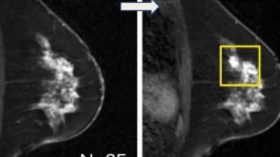
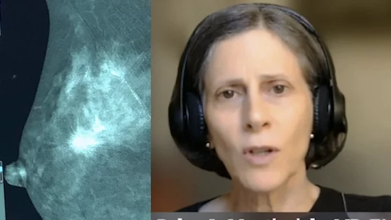
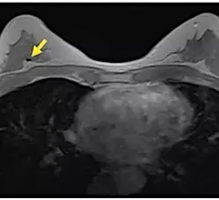
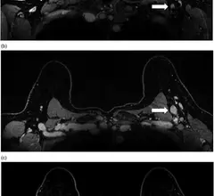
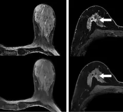
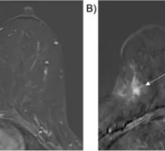
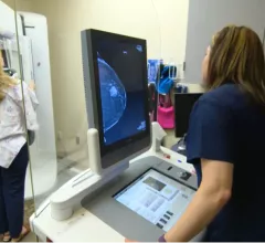
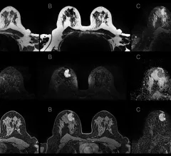
Magnetic resonance imaging of the breast (breast MRI), commonly used for dense breasts, is a highly accurate imaging modality for detecting early breast cancer and other abnormalities without using excess radiation. It’s often used alongside digital mammograms and digital breast tomosynthesis to offer detailed visualization of breast lesion margins. 

A mammography exam in progress with the patient on the breast imaging system and the technologist acquiring images. The image on the screen shows a suspect lesion.

4 key trends in breast imaging

These trends include growth in 3D mammography, supplemental imaging for women with dense breasts and in the role of artificial intelligence.

January 31, 2023
Example of various breast MRI protocol sequences that offer different types of soft tissue enhancement. Imaging performed on a Siemens Magnetom system. Breast MRI can help see through dense breast tissue to better detect cancers. #densebreasts #Breastdensity #BreastMRI

MRI a 'far superior' modality for dense breast imaging

The modality outperformed digital breast tomosynthesis, handheld ultrasound and automated breast ultrasound for cancer detection in a large cohort of women with dense breast tissue.

January 31, 2023
womanconsultation.jpg

ACR recommendations result in 15-fold increase in breast MRI eligibility among cancer survivors

Prior to 2018, it was recommended that women at higher-than-average risk undergo supplemental breast MRI screening only if they had a personal history of breast cancer in addition to carrying a hereditary breast cancer gene mutation.

December 19, 2022
Women with dense breasts and benign breast disease have increased risks of developing #breastcancer

Many providers remain underinformed about increased cancer risks associated with breast density

JoAnn Pushkin, breast cancer survivor and executive director of DenseBreast-info.org, sounded the alarm on the lack of awareness surrounding breast density and its associated cancer risks during a presentation at this year’s annual RSNA meeting.

November 29, 2022
synthetic contrast-enhanced breast MRI

Are synthetic contrast-enhanced breast MRI images as good as the real thing?

Synthesizing contrast-enhanced imaging from pre-contrast inputs using deep learning can help overcome obstacles involved in the use of GBCAs.

November 18, 2022
womanconsultation.jpg

Personalized breast cancer risk assessment could reduce overdiagnosis

Personalized breast cancer screening programs could help women better understand their personal cancer risks and guide providers in recommending mammographic assessments based on these risks. 

November 16, 2022
Constance "Connie" Lehman, MD, PhD,, chief of breast imaging, co-director of the Avon Comprehensive Breast Evaluation Center at the Massachusetts General Hospital, and professor of radiology at Harvard Medical School, discusses trends in breast imaging.

VIDEO: Connie Lehman discusses trends in breast imaging

Connie Lehman, MD, PhD, chief of breast imaging, co-director of the Avon Comprehensive Breast Evaluation Center at the Massachusetts General Hospital, and professor of radiology at Harvard Medical School, discusses trends she sees in breast imaging.

October 24, 2022
Contrast-enhanced mammography uses iodine contrast injected into a patient and mammography system to image contrast uptake or areas of increased vascular activity, which is typical of cancers. This can help image through dense breast tissue to find cancers that are otherwise masked by dense breast tissue.

VIDEO: Why contrast enhance mammography might be the ideal supplemental imaging

Connie Lehman, MD, PhD, chief of breast imaging, co-director of the Avon Comprehensive Breast Evaluation Center at the Massachusetts General Hospital, explains why she thinks contrast-enhanced mammography will likely become the go-to supplemental imaging modality for women with dense breasts.

October 20, 2022

Around the web

Automated AI-generated measurements combined with annotated CT images can improve treatment planning and help referring physicians and patients better understand their disease, explained Sarah Jane Rinehart, MD, director of cardiac imaging with Charleston Area Medical Center.

Two advanced algorithms—one for CAC scores and another for segmenting cardiac chamber volumes—outperformed radiologists when assessing low-dose chest CT scans. 

"Gen AI can help tackle repetitive tasks and provide insights into massive datasets, saving valuable time," Thomas Kurian, CEO of Google Cloud, said Tuesday. 

Trimed Popup
Trimed Popup