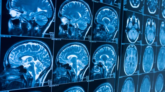
MRI study shows brain changes linger 6 months after concussion
Using a technique that combines two MRI scans, researchers have developed a new, objective way to monitor concussions, according to a study published in NeuroImage: Clinical.
“Diagnosis of concussion is subjective right now,” said senior author Ravi Menon, PhD, of Western University, London, in a statement. “There is a long checklist that trained physicians can look at, and while it is pretty good at diagnosing the initial concussion, it is not sensitive to the longer-term brain changes and making decisions about when someone is okay to return to play.”
Over five years, a total of 52 female rugby players, including 21 with concussions, underwent both fMRI and diffusion tensor imaging (DTI) throughout the season and during the postseason. By analyzing each scan separately, and comparing them to the scans of healthy volunteers, Menon et al. identified a new, and perhaps more importantly, objective way to monitor brain injuries.
They found three unique brain signatures: One that reveals acute brain changes after a concussion; a second that identifies persistent changes six months following a concussion; and one signature that shows evidence of a past concussion.
According to Menon et al. their study aligns with increasing evidence that shows functional and structural brain changes can remain six months after a concussion.
"We were able to show evidence of prior concussion history through this method,” Menon said in the same statement. “This component correlates directly with the number of previous concussions that an athlete has had. This hasn’t been shown before.”

