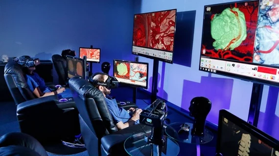
How virtual reality is taking surgical training to the next level
At Stanford University Medical School in California, virtual reality is helping to make surgical training and planning more efficient and patient-centered all while reframing education for medical students, according to an article published online Jan. 9 by Fortune.
Virtual reality can be one of two experiences: either fully immersive in which the users only see a computer-generated environment or mixed/augmented reality where 3D images such as MRIs or CT scans are projected onto patients or physical surroundings.
Because medical images become so immersive and intricately detailed when using virtual reality, medical students who use the technology are graded on whether they make a mistake in the virtual operating room instead of how long it takes them to perform a procedure, according to the article.
This is the case at Stanford’s Neurosurgical Simulation and Virtual Reality Center, which resembles a miniature movie theater where students and surgeons can use virtual reality while spectators watch on large TV screens mounted on the wall. The center, which opened, in 2016 has allowed more than 400 neurosurgery patients to see their surgeries before having their procedures.
“They can immerse themselves in their brain,” Gary Steinberg, MD, PhD, chair of neurosurgery at Standford University School of Medicine and co-creator of the Neurosurgical Simulation and Virtual Reality Center, told Fortune. “It puts them at ease and shows them exactly what we’re going to do.”
See the entire article below.

