Radiologist uses virtual reality to improve thyroid nodule treatment
A University of Virginia Health interventional radiologist is using virtual reality to improve the way doctors care for patients with thyroid nodules.
Thanks in part to a grant from UVA’s Office of Graduate Medical Education, Ziv Haskal, MD, developed an 11-minute virtual reality tool which offers a better way for physicians to learn about a less-invasive treatment for thyroid patients. It’s part of a larger project to develop educational materials for the entire university.
“The VR experience uniquely places the viewer into the room right next to the operator,” Haskal said in a UVA news release. “I want interventional radiologists to say, ‘I can see myself doing this.’”
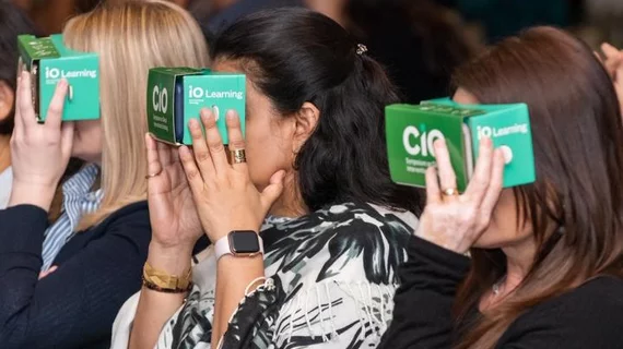
Nearly 200 physicians got to test out the experience when Haskal debuted his educational video at the Symposium on Clinical Interventional Oncology in Miami, which wrapped up on Oct. 13.
Those doctors wearing VR headsets were taught about thermal ablation, a less invasive procedure that uses a heat-packed probe to shrink cancerous lumps in the thyroid. The method has shorter recovery times, less scarring and is more apt to preserve thyroid function compared to open surgery. It’s currently utilized more outside the U.S., where it isn’t usually covered by insurance.
Despite this, Haskal believes that his VR creation, along with other educational offerings, can make UVA a training center for thermal ablation.
“This technology has incredible potential to improve care, whether it is by better training doctors to perform procedures or helping patients know what to expect when they arrive at the hospital,” Haskal said.

