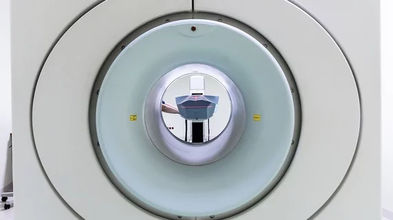
Researchers develop a ‘significant breakthrough’ in magnetic resonance imaging
Scientists working across multiple fields of research have developed a new hyperpolarization technique that they say can significantly change the field of magnetic resonance imaging.
The proposed concept involves rapidly hyperpolarizing fumarate, a key metabolic product in human energy creation, using parahydrogen, experts explained in the Proceedings of the National Academy of Sciences. It’s a highly scientific process, but one that could more accurately image kidney injuries, the effects of a heart attack, and much more.
Additionally, parahydrogen-induced polarization, or PHIP, is much easier and more cost-effective compared to the gold standard for hyperpolarizing fumarate.
"We have made a significant breakthrough as our approach is not only cheap but also fast and easy to handle," project leader James Ellis, PhD, of Johannes Gutenberg University Mainz in Germany, explained on Tuesday.
The benefits of MRI are widely known, yet the technique is essentially limited to imaging water molecules in the body, Ellis explained. And because of this, researchers are always working on ways to enhance the technology behind this modality.
While their PHIP approach is a promising biomarker for imaging how the body creates and uses energy, the group did encounter a few problems.
For one, there are a large number of unwanted chemical contaminants that must be removed from the final substance before it can be injected into humans like an imaging tracer.
Ellis and co-investigators did find success purifying the hyperpolarized fumarate, however, which creates a product without toxic substances seemingly safe for humans.
Finally, the researchers noted that preclinical studies have shown this imaging technique is suitable for monitoring how cancerous tumors respond to therapy along with a host of other healthcare advances.
Others who helped contribute to this study included: Technical University Darmstadt and Kaiserslautern, both in Germany, the University of California Berkeley, the University of Turin in Italy, and the University of Southampton in England.

