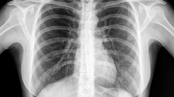
Deep learning models predict COPD survival based on chest radiographs
Predicting prognoses for patients with chronic obstructive pulmonary disease could become a much simpler process with the assistance of deep learning.
That’s according to a new paper published in Radiology June 7. In it, experts explored the efficacy of two deep learning models for grading COPD severity based on chest radiographs—a method that has not yet been thoroughly tested, the authors noted.
“Chest radiography is the most widely performed radiologic examination, but its role as a prognostic predictor has not been thoroughly investigated. Although some chest radiographic features, such as hyperinflation or degree of emphysema, are known to reflect pulmonary function, those findings are difficult to quantify, and thus have seldom been used in previous survival prediction models,” corresponding author Chang Min Park, of the Department of Radiology at Seoul National University Hospital, and co-authors explained.
Currently, predicting COPD prognosis involves complex assessments and exercise tests, which is impractical on many fronts, the experts said. The application of deep learning models to chest radiographs could save patients time and money and help providers to develop COPD management plans specific to patients’ needs based on their disease severity.
To test their hypothesis, the researchers developed two deep learning models using data from patients who had completed both postbronchodilator spirometry and chest radiographs between 2011 and 2015. The data were split into training, validation and internal test sets and used to train the models to predict the 5-year survival of COPD patients.
The model that used chest radiographs to predict survival performed better than the model that integrated age, body mass index, and forced expiratory volume in 1 second (FEV1%). That same model achieved an AUC of .73, .67 and .76 in three external test sets and was observed to be as reliable as other commonly used COPD-related clinical indexes and scores, such as BODE, CAT and SGRQ.
The authors noted an additional benefit of their prediction models is that they incorporated inputs that are easily accessible, such as chest x-rays and FEV1%, both of which are easy to obtain on follow-up visits.
To view the full study, click here.
Related deep learning and artificial intelligence news:
Diagnosing osteoporosis using a deep-radiomics-based approach
Deep learning algorithm predicts emphysema mortality
DL networks augment radiologist performance for thyroid cancer detection
Stratifying patients by risk of poor outcomes could reduce overtreatment of lung cancer
References:
Deep Learning Prediction of Survival in Patients with Chronic Obstructive Pulmonary Disease Using Chest Radiographs. Ju Gang Nam, Hye-Rin Kang, Sang Min Lee, Hyungjin Kim, Chanyoung Rhee, Jin Mo Goo, Yeon-Mok Oh, Chang-Hoon Lee, and Chang Min Park. Radiology.

