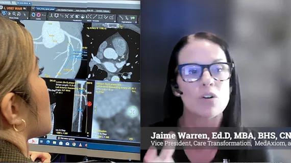
Jaime Warren, EdD, FACC, vice president of care transformation at MedAxiom, an ACC company, explains what is needed to address staffing shortages created by a 94% increase in cardiac CT volumes over the past 5 years.
After several radiology patients were sickened following an injection of iodinated contrast, the hospital where the incidents occurred has revealed the culprit.
The Symphony Thrombectomy System eliminates tradeoffs between two priorities—leveraging large-bore power and ease of use vs. efficiently reducing clot burden and delivering improved speed.
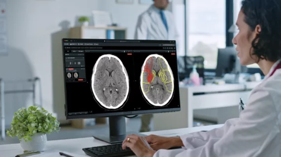
This week, health officials in the United Kingdom shared how an artificial intelligence-enabled platform has drastically improved stroke outcomes in the region.
NIH is hopeful the research will enable doctors to detect signs of stroke damage earlier, opening the door for them to initiate treatments in a timelier manner.
Leqembi (lecanemab) was approved by the FDA in 2023. It is an infusion drug that has been shown to reduce Alzheimer’s-related cognitive decline by up to 27%.
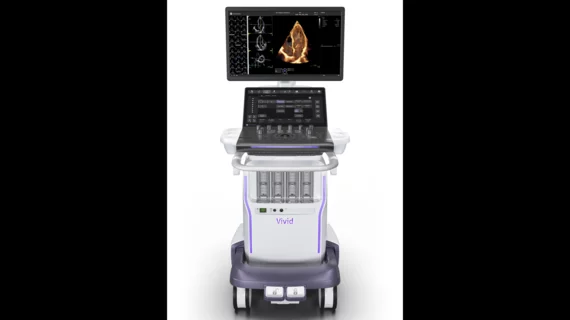
The company describes Vivid Pioneer as its “most advanced, ultra-premium and adaptive cardiovascular ultrasound system yet.” It includes new and improved AI capabilities and a compact design that is still fully functional in tight workspaces.