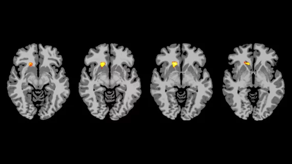
AI, brain MRI combine to improve detection of ADHD
A new deep learning approach can boost the performance of brain MRI to better predict attention deficit hyperactivity disorder (ADHD), according to a Dec. 11 study published in Radiology: Artificial Intelligence.
More than 6.1 million children were diagnosed with ADHD in 2016. Despite these numbers, there is no single test or imaging exam that can confidently diagnose a patient; instead, the disorder is determined from a series of symptoms and behavior-based tests.
The new AI-based technique utilizes a multi-scale approach to analyze many brain connectomes for a more comprehensive view of the organ. Connectomes are built using spatial regions across MRI scans, which are called parcellations, and are believed to breakdown in patients with ADHD. Parcellations are grouped according to brain function, anatomy or both.
"Our results emphasize the predictive power of the brain connectome," senior author Lili He, PhD, from the Cincinnati Children's Hospital Medical Center, said in a statement. "The constructed brain functional connectome that spans multiple scales provides supplementary information for the depicting of networks across the entire brain."
In order to build their deep learning model, the team collected data from the NeuroBureau ADHD-200 dataset. Within that set is brain connectome information from 973 patients, along with their personal characteristics, such as gender and IQ.
Compared to a single-scale method, the multi-scale AI-based approach was “significantly” better, using multiple connectome maps based off of numerous brain parcellations.
He and colleagues even believe the technique could apply to conditions outside of ADHD.
"This model can be generalized to other neurological deficiencies," she said in the statement. "We already use it to predict cognitive deficiency in pre-term infants. We scan them soon after birth to predict neurodevelopmental outcomes at two years of age."
Beyond that, He et al. plan to improve their model with larger neuroimaging datasets and deepen their understanding of why breakdowns in connectomes are associated with ADHD.

