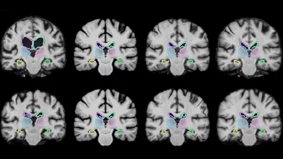
MIT-developed AI algorithm compares 3D images 1,000 times faster than standard techniques
MIT researchers have developed an artificial intelligence-based algorithm that can register 3D images 1,000 times more quickly than standard medical image registration techniques.
The algorithm, called VoxelMorph, uses machine learning technology to register thousands of pairs of images to gain information on how to correctly align them and compare anatomical differences for diagnostic purposes in a matter of minutes, according to a June 18 news release by MIT.
“The tasks of aligning a brain MRI shouldn’t be that different when you’re aligning one pair of brain MRIs or another,” said co-author Guha Balakrishnan, a graduate student in MIT’s Computer Science and Artificial Intelligence Laboratory and department of engineering and computer science, in a prepared statement. “There is information you should be able to carry over in how you do the alignment. If you’re able to learn something from previous image registration, you can do a new task much faster and with the same accuracy.”
VoxelMorph is powered by a convolutional neural network (CNN), a machine-learning approach commonly used for image processing.
Once the algorithm is trained, Balakrishnan and colleagues explained that the alignment parameters of the images obtained are used to map all the pixels of one image to another simultaneously. It's also able to easily match scans coming from different imaging machines with different spatial orientations.
"Importantly, the algorithm is 'unsupervised', meaning it doesn’t require additional information beyond image data," according to the news release. "Some registration algorithms incorporate CNN models but require a 'ground truth', meaning another traditional algorithm is first run to compute accurate registrations; the researchers’ algorithm maintains its accuracy without that data."
Although the findings only include using the algorithm to analyze brain scans, the researchers explained that VoxelMorph can analyze of wide range of images and has the potential to register scans in near real-time for surgeons during an operation.
The researchers' findings are published in two papers that will be presented at the International Conference on Computer Vision and Pattern Recognition this week in Salt Lake City, Utah.

