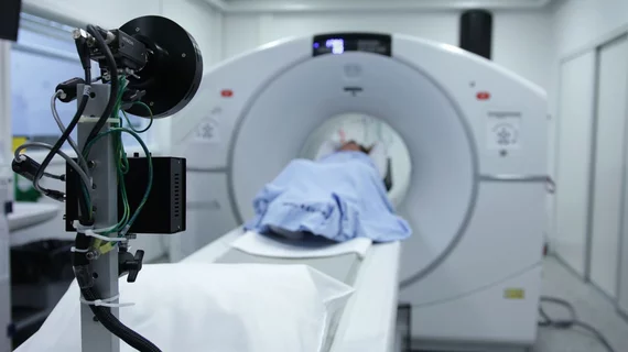
We can now diagnose Alzheimer's using only an MRI and an algorithm
With the help of machine learning, a simple MRI of the brain can diagnose Alzheimer’s Disease in its early stages when the neurological condition has been historically difficult to identify on imaging.
Funded by the National Institute for Health and Care Research (NIHR) Imperial Biomedical Research Center, new work shared in Communications Medicine highlights how experts were able to develop and train an algorithm to detect the presence of Alzheimer’s Disease on a simple MRI exam. After dividing the brain into 115 different regions, 660 size, shape and texture imaging features were used to assess neurological changes related to Alzheimer’s. Encouragingly, the scans did not require the latest equipment advances and were conducted on a standard machine that is readily available at most hospitals—a 1.5 Tesla.
“Although neuroradiologists already interpret MRI scans to help diagnose Alzheimer’s, there are likely to be features of the scans that aren’t visible, even to specialists. Using an algorithm able to select texture and subtle structural features in the brain that are affected by Alzheimer’s could really enhance the information we can gain from standard imaging techniques,” explained Dr. Paresh Malhotra, who is a consultant neurologist at Imperial College Healthcare NHS Trust and a researcher in Imperial’s Department of Brain Sciences.
Experts used the machine learning-based method on 400 brain MRIs of patients with early or late-stage Alzheimer’s, in addition to scans of healthy controls and individuals with other neurological conditions. The algorithm yielded an impressive accuracy of 98% for predicting whether patients had Alzheimer’s or not. This, experts involved in the study shared, is “an important step forward.”
“Currently no other simple and widely available methods can predict Alzheimer’s disease with this level of accuracy,” said Professor Eric Aboagye, from Imperial’s Department of Surgery and Cancer, who led the research. “Many patients who present with Alzheimer’s at memory clinics do also have other neurological conditions, but even within this group our system could pick out those patients who had Alzheimer’s from those who did not.”
The machine-learning based method was also able to distinguish between early and late-stage Alzheimer’s in 78% of cases, according to the research. This was achieved, in part, by identifying changes in areas of the brain that were not previously associated with Alzheimer’s, such as the cerebellum and the ventral diencephalon.
Researchers involved in the study suggested that their work could help streamline the process of receiving a diagnosis and beginning treatment and/or management in the future, in addition to potentially expanding research into new areas of the brain that could offer added insight into how the condition manifests.
Related Alzheimer's content:
New advanced PET imaging reveals root of cognitive decline in patients with Alzheimer's
CMS coverage decision for Alzheimer's drug, related PET scans sparks concern in imaging community
University awarded research grant to study Alzheimer's using specialized brain MRI

