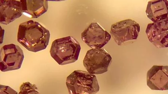
Diamond tracers may be vital key to cost-friendly, high-resolution imaging
Microscopic diamond tracers can be detected simultaneously via two distinct imaging approaches—optical and MRI—to produce higher-quality images in a fraction of the time, according to recently published research.
Traditional microscopes utilize optical fluorescence to produce high-resolution images but lack the ability to image deep into tissue. MR imaging, on the other hand, uses radiofrequency that can reach everywhere in the body yet produces lower-resolution scans.
With hyperpolarized biological diamond tracers, however, both can be achieved at once to capture clear images up to 10 times deeper than using light-based techniques alone, one University of California, Berkeley researcher proved in a study published in the journal Proceedings of the National Academy of Sciences.
"This is perhaps the first demonstration that the same object can be imaged in optics and hyperpolarized MRI simultaneously," Ashok Ajoy, UC Berkeley assistant professor of chemistry, explained in a university news item published May 17. "There is a lot of information you can get in combination, because the two modes are better than the sum of their parts.”
These diamond particles are relatively new but have been investigated by Ajoy in the past. Together with experts from the U.S. Department of Energy, Ajoy published research in 2018 suggesting defects in nanoscale and microscale diamonds could eliminate the need for costly, bulky magnets used in MRI and nuclear magnetic resonance systems.
In this latest experiment, the Berkeley assistant professor used a magnetic field equal to that of a cheap refrigerator magnet to hyperpolarize carbon-13 atoms within microdiamonds.
"It turns out that if you shine light on these particles, you can align their spins to a very, very high degree—about three to four orders of magnitude higher than the alignment of spins in an MRI machine," Ajoy said. "Compared to conventional hospital MRIs, which use a magnetic field of 1.5 teslas, the carbons are polarized effectively like they were in a 1,000-tesla magnetic field."
Given these diamond tracers are relatively cheap and easy to handle, Ajoy said his technique could one day bring advanced imaging rivaling MRI to hospitals that can’t afford the high price tag of magnetic resonance machines.

