Artificial Intelligence

Artificial intelligence (AI) is becoming a crucial component of healthcare to help augment physicians and make them more efficient. In medical imaging, it is helping radiologists more efficiently manage PACS worklists, enable structured reporting, auto detect injuries and diseases, and to pull in relevant prior exams and patient data. In cardiology, AI is helping automate tasks and measurements on imaging and in reporting systems, guides novice echo users to improve imaging and accuracy, and can risk stratify patients. AI includes deep learning algorithms, machine learning, computer-aided detection (CAD) systems, and convolutional neural networks. 

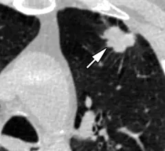
AI beats standard regression models at predicting lung cancer risk

Not all AI or regression models are the same, nor do they all incorporate the same data when assessing patient risk.

AI reduces CT lung cancer screening workload by nearly 80%

And it can do so with almost 100% accuracy as a first reader, according to a new large-scale analysis.

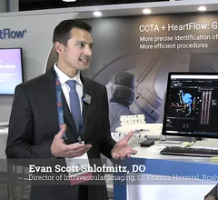
Evan Scott Shlofmitz, DO, Director of Intravascular Imaging, St. Francis Hospital, in Roslyn, New York, explains how he uses Heartflow's artificial intelligence technology to assess a patient's coronary artery disease from noninvasive CT scans to preplan PCI procedures.

How AI and CCTA help heart teams plan ahead before PCI

Evan Shlofmitz, DO, director of intravascular imaging at St. Francis Hospital, explains how advanced artificial intelligence technology is used to assess a patient's CT scan before they undergo PCI.

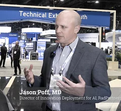
Jason Poff, MD, director of innovation deployment for artificial intelligence (AI) at RadPartners, explains the five-step process he uses to evaluate medical imaging AI.

5 steps for evaluating radiology AI applications

Jason Poff, MD, director of innovation deployment for artificial intelligence at Radiology Partners, explains the process he uses to evaluate medical imaging AI. 
 

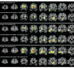
AI detects subtle changes in images over time.

Adaptable AI system detects subtle changes in imaging, has potential across multiple clinical settings

The Learning-based Inference of Longitudinal imAge Changes, or LILAC, system harnesses machine learning to review medical images that have been collected over a prolonged period.

Thumbnail

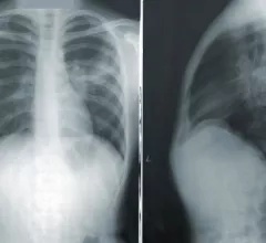
New algorithm models radiologists' eye movements to interpret chest X-rays

The algorithm has an edge over standard black box-style artificial intelligence applications because providers are able to see how it reaches conclusions.

Video of James Min, MD, explaining the future of cardiac care using CT and AI plaque analysis to create a personalized and more accurate cardiac risk assessment, similar to a mammogram for the heart.

Embracing the future: James Min left academia to push for a paradigm shift in preventive cardiology

James Min, MD, Cleerly's founder and CEO, changed careers to address what he saw as a major unmet need in cardiology.

thyroid biopsy

Risk prediction algorithm slashes number of unnecessary thyroid nodule biopsies

Although the vast majority of nodules are benign, many are referred for biopsy as a precaution to rule out malignancy.

Around the web

GE HealthCare designed the new-look Revolution Vibe CT scanner to help hospitals and health systems embrace CCTA and improve overall efficiency.

Clinicians have been using HeartSee to diagnose and treat coronary artery disease since the technology first debuted back in 2018. These latest updates, set to roll out to existing users, are designed to improve diagnostic performance and user access.

The cardiac technologies clinicians use for CVD evaluations have changed significantly in recent years, according to a new analysis of CMS data. While some modalities are on the rise, others are being utilized much less than ever before.