Artificial Intelligence

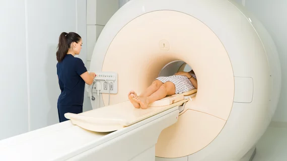
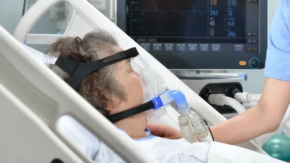
Artificial intelligence (AI) is becoming a crucial component of healthcare to help augment physicians and make them more efficient. In medical imaging, it is helping radiologists more efficiently manage PACS worklists, enable structured reporting, auto detect injuries and diseases, and to pull in relevant prior exams and patient data. In cardiology, AI is helping automate tasks and measurements on imaging and in reporting systems, guides novice echo users to improve imaging and accuracy, and can risk stratify patients. AI includes deep learning algorithms, machine learning, computer-aided detection (CAD) systems, and convolutional neural networks. 

Natural language processing tool data mines unstructured neuroradiology reports

 Diagnostic radiology generates a lot of data. Although the practice’simages can be parsed and relevant information can be acquired via theutilization of DICOM tools, free-text generated radiology reports havelong languished in silos of unconnected data. A natural languageprocessing (NLP) application developed at Massachusetts GeneralHospital (MGH) in Boston holds the promise of allowing researchers thecapability to data mine unstructured radiology reports.

Worklist sharing adds balance to radiologist reading load

 The average diagnostic image reading volume of a radiologist working inthe United States, according to a disparate variety of sources, isbetween 17,000 to 20,000 or more studies annually—depending on theprocedure mix. Outliers exist on both ends of the spectrum, and chestx-rays are easier to clear than a mammogram or neurological MRI study;however, it is clear that radiology groups are confronting anincreasing number of exams each year.

Emageon names Perkins as COO

Emageon, a provider of medical IT systems forhospitals and healthcare networks, has named Chris E. Perkins as chiefoperating officer (COO).

MedcomSoft appoints former Bush staffer to board

MedcomSoft, an EHR provider, has announced that James Haveman has joined its healthcare advisory board. 

Health IT forum convenes to decide fate of Massachusetts EMRs

Dec. 6 – Massachusetts must decide quickly how to pay for the newelectronic medical records systems and what role government shouldplay, according to healthcare officials who attended the Mass HealthPolicy Forum in Boston on Wednesday, reported The Boston Globe.

Study shows superior sensitivity for 3D virtual colonography

 Primary interpretation of diagnostic images is traditionally conductedin two-dimensional views. Although interpretative viewing in 2D planeshas historically demonstrated excellent results, primary evaluation ofCT colonography exams is best conducted in 3D view, according to arecent study published in the American Journal of Roentgenology.

Visual probe localization enables cost-effective 3D ultrasound

 A team from the Multimedia University, Cyberjava, in Selangor,Malaysia, has developed a method of creating 3D ultrasound images usinga 2D ultrasound probe with a conventional digital camera. Theirtechnique, described in the December issue of the Journal of Digital Imaging,holds the promise to allow clinicians in cash-strapped practices toconduct modified 3D ultrasound exams with their current equipment.

Advanced Visualization: More Uses, More Confidence

Post-processing software allows clinicians to better track cancertherapies, plan surgeries, and diagnose heart, brain and intestinaldisease. Newer tools let users subtract unnecessary structures,allowing for better evaluation of problem areas. Use of and uses forthe software will only increase as data sets grow and new applicationsare added to the list.

Around the web

Positron, a New York-based nuclear imaging company, will now provide Upbeat Cardiology Solutions with advanced PET/CT systems and services. 

The nuclear imaging isotope shortage of molybdenum-99 may be over now that the sidelined reactor is restarting. ASNC's president says PET and new SPECT technologies helped cardiac imaging labs better weather the storm.

CMS has more than doubled the CCTA payment rate from $175 to $357.13. The move, expected to have a significant impact on the utilization of cardiac CT, received immediate praise from imaging specialists.