Echocardiography

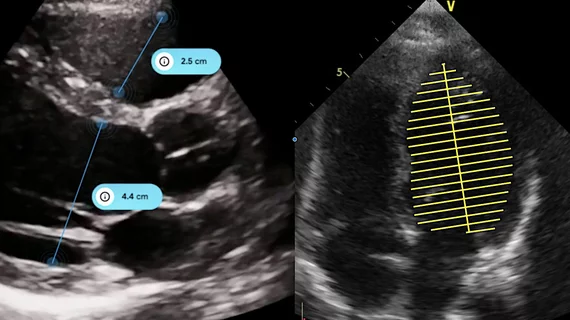
Cardiac ultrasound uses reflected sound waves (echos) to create images of anatomy inside the body. Echocardiograms are the primary cardiac imaging modality used to assess the heart and diagnose or track cardiac issues. Echo is the gold standard imaging modality to assess the heart, particularly with calculating left ventricular ejection fraction (LVEF), which is a measure of cardiac output. In addition to noninvasive standard transthoracic echo (TTE), invasive transesophgeal echo (TEE) is also used when clearer, more detailed imaging of the heart is needed. Both 3D and 4D echo echo systems are rapidly gaining wider adoption and enable new types of assessments, especially in the structural heart space and in transcatheter procedural guidance. Find news on general ultrasound imaging.
Cardiac imaging pioneer remembered for his joy and enthusiasm

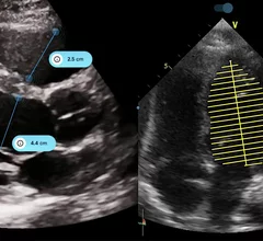
Former American Society of Echocardiography president and well-known cardiac ultrasound pioneer Roberto Lang, MD, died at the age of 73. He helped develop 3D echo technology that is now used by care teams on a daily basis.

ROB’E Robotic transesophageal echocardiography (TEE)

Cardiologists make history, use new robotic imaging system for first time

Researchers believe a new robotic TEE system could improve the long-term health of interventional echocardiographers in addition to several other key benefits.

GE HealthCare Optison ultrasound enhancing agent

FDA approves popular ultrasound enhancing agent for pediatric patients

Unlike other UEA options, GE HealthCare's Optison does not contain polyethylene glycol. The FDA approved its use for adult patients back in 1997.

HeartFocus DESKi

FDA clears AI-powered echocardiography software

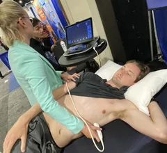
HeartFocus guides users through the entire process, making it so that even novices can deliver high-quality echocardiograms every time.

The Agitation Solutions Orbis Microbubble Generator simplifies and streamlines the process of saline agitation and delivery for cardiac bubble studies using a single use syringe. It is was cleared for use in Canada in 2024.

Right heart cardiac ultrasound contrast agent for shunts enters U.S. trial

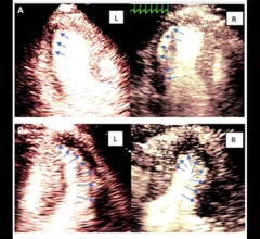
Currently there are no FDA cleared echocardiography contrast agents commercially available for use on the right side of the heart to detect shunts, but a new agent that promises to improve workflow and imaging is about to enter a U.S. trial. 

Key trends in diagnostic heart testing: CT on the rise as some traditional techniques fall out of favor

The cardiac technologies clinicians use for CVD evaluations have changed significantly in recent years, according to a new analysis of CMS data. While some modalities are on the rise, others are being utilized much less than ever before.

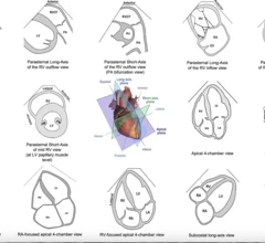
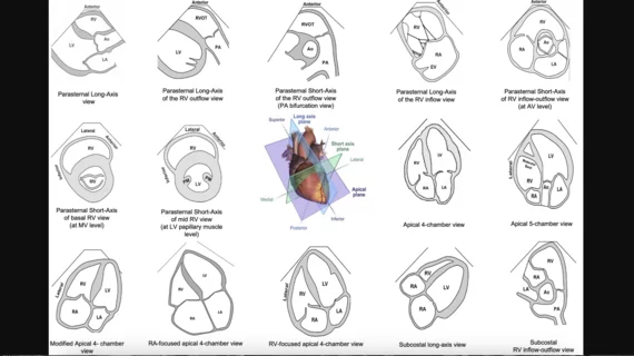
Standard transthoracic imaging planes for comprehensive right heart assessment. Several key views should be included in the standard echocardiographic assessment of the right heart. Image and caption courtesy of the American Society of Echocardiography.

ASE updates recommendations for assessing right heart function in patients with pulmonary hypertension

The new guidelines were designed to ensure sonographers and other members of the heart team have the information they need to screen patients when appropriate and identify early warnings signs of PH. 

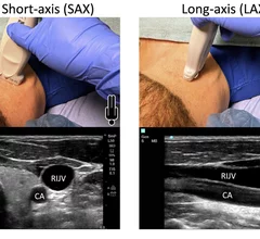
Figure from the new ASE guideline for ultrasound guided vascular access, showing vessel and probe orientation on the transverse plane short axis (SAX) view perpendicular to the vessels, or in longitudinal long axis plane (LAX) coaxial to the vessels. Image courtesy of ASE

ASE shares new guidelines for ultrasound-guided vascular access

The American Society of Echocardiography included descriptions, diagrams and ultrasound images in the new document to make it as helpful as possible. 

Around the web

Former American Society of Echocardiography president and well-known cardiac ultrasound pioneer Roberto Lang, MD, died at the age of 73. He helped develop 3D echo technology that is now used by care teams on a daily basis.

Imaging and radiology are in a transition right now as more departments and practices are choosing to bring their 3D labs in-house.  

John Simon, MD, CEO of SimonMed Imaging, says imaging has considerably advanced for noninvasive detection of disease and it may be time for it to play a greater role in annual physicals, especially in executive physical exams.