Echocardiography

Cardiac ultrasound uses reflected sound waves (echos) to create images of anatomy inside the body. Echocardiograms are the primary cardiac imaging modality used to assess the heart and diagnose or track cardiac issues. Echo is the gold standard imaging modality to assess the heart, particularly with calculating left ventricular ejection fraction (LVEF), which is a measure of cardiac output. In addition to noninvasive standard transthoracic echo (TTE), invasive transesophgeal echo (TEE) is also used when clearer, more detailed imaging of the heart is needed. Both 3D and 4D echo echo systems are rapidly gaining wider adoption and enable new types of assessments, especially in the structural heart space and in transcatheter procedural guidance. Find news on general ultrasound imaging.
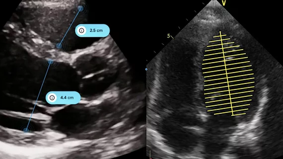
Example of the Siemens Origin AI-automated cardiac ultrasound system performing auto contours and measurements after the AI sees what is being imaged and the operator hits the AI button on the console. The system is designed to be an assistant to the operator and knows the next steps in the exams.

Siemens Healthineers unveils 'truly revolutionary' AI-enabled echo system

The new-look system is able to evaluate a situation and anticipate what users may need next.

Video of ASE President Stephen Little, MD, discussing trends in interventional echo at the ASE 2023 annual meeting.

Interventional echocardiography expected to grow thanks to new structural heart procedures

American Society of Echocardiography (ASE) President Stephen Little, MD, says there will be an increasing need for interventional echocardiographers in the near future.

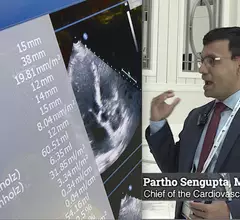
Video of Partho Sengupta, MD, explaining two major shifts in AI for echocardiography at ASE 2023. These include GPT and deep learning to automate measurements. #ASE23 #ASE2023 #ASE #AI

2 key reasons the use of AI in echocardiography is growing

Partho Sengupta, MD, an expert on the use of AI in cardiology, says some of the technologies being developed today will fundamentally change how cardiologists and sonographers do their jobs.

HCM expert Sherif F. Nagueh, MD, FACC, FASE, professor of cardiology, Methodist DeBakey Heart and Vascular Center, explains the key points of HCM imaging. #ASE #HCM

How medical imaging helps cardiologists treat hypertrophic cardiomyopathy

Hypertrophic cardiomyopathy expert Sherif F. Nagueh, MD, examined imaging's role in treating this serious condition. 

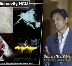
Hari Naidu, MD, explains that inconsistent diagnostic imaging and how it is reported has likely led to a major underdiagnosis of HCM. #ASE #ASE2023 #HCM

Why hypertrophic cardiomyopathy may be widely underdiagnosed

One expert believes the lack of standardization for how diagnostic imaging in conducted and reported has led to a major underdiagnosis of HCM.

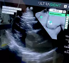
FDA clears new AI to guide point-of-care cardiac ultrasound exams

The FDA approved UltraSight's AI to show ultrasound users how to perform a diagnostic quality cardiac ultrasound exam at the point of care in multiple settings.

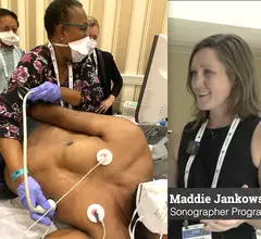
Video of Maddie Jankowski explaining sonographer highlights and trends at the American Society of Echo (ASE) 2023 meeting.

The rise of AI and other key takeaways for sonographers at ASE 2023

Cardiac sonographer Maddie Jankowski, BS, ACS, RDCS, joined us to explore key trends and updates for sonographers from ASE 2023 in National Harbor, Maryland. 

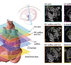
Axial planes suggested for screening the fetal heart at the time of the obstetric anatomic survey and as an initial series obtained during fetal echocardiography. #Fetalecho

ASE updates fetal echocardiography guidelines

The new 44-page document focuses on the detection, classification and risk assessment of congenital heart issues.

Around the web

The new technology shows early potential to make a significant impact on imaging workflows and patient care. 

Richard Heller III, MD, RSNA board member and senior VP of policy at Radiology Partners, offers an overview of policies in Congress that are directly impacting imaging.
 

The two companies aim to improve patient access to high-quality MRI scans by combining their artificial intelligence capabilities.