Echocardiography

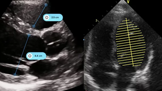
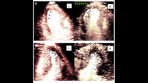
Cardiac ultrasound uses reflected sound waves (echos) to create images of anatomy inside the body. Echocardiograms are the primary cardiac imaging modality used to assess the heart and diagnose or track cardiac issues. Echo is the gold standard imaging modality to assess the heart, particularly with calculating left ventricular ejection fraction (LVEF), which is a measure of cardiac output. In addition to noninvasive standard transthoracic echo (TTE), invasive transesophgeal echo (TEE) is also used when clearer, more detailed imaging of the heart is needed. Both 3D and 4D echo echo systems are rapidly gaining wider adoption and enable new types of assessments, especially in the structural heart space and in transcatheter procedural guidance. Find news on general ultrasound imaging.

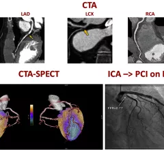
A figure from the 2022 CAD non-invasive imaging guidelines showing a comparison of computed tomography angiography (CTA) and a SPECT-CT vs. an invasive angiogram from the cath lab showing the same blockage in a coronary artery.

New multi-society recommendations highlight role of non-invasive imaging in evaluating coronary artery disease

A new, multi-society document, "Non-Invasive Imaging in Coronary Syndromes," focuses on how multiple imaging techniques can evaluate different aspects of coronary artery disease (CAD), all without the need for invasive angiograms.

William A. Zoghbi, MD, MACC, FAHA, FASE, is the chair of the Department of Cardiology at the Houston Methodist DeBakey Heart and Vascular Center, and past president of both the American College of Cardiology (ACC) and the American Society of Echocardiography (ASE).

VIDEO: New advances in echocardiography

William A. Zoghbi, MD, past president of the ACC and ASE, discussed the latest trends in cardiac ultrasound technology. 

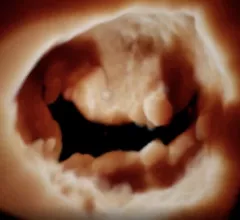
The mitral valve seen on 3D cardiac ultrasound offers a noninvasive surgeon's view of the valve for better diagnostics and pre-planning of structural heart procedures. Example from the GE Vivid E95 FlexiLight photo-realistic 3D/4D lighting system.

Q&A: William Zoghbi breaks down the future of echocardiography

In a new interview, former ACC and ASE President William A. Zoghbi, MD, reviews some of the biggest trends in cardiac ultrasound.

Thumbnail

Medtech company Us2.ai gains FDA clearance for automated echocardiography software

The news comes following a validation study at Brigham and Women's Hospital and multiple trials with pharmaceutical partners.

Thumbnail

Echocardiogram add-on predicts patients at risk of developing heart problems from COVID-19

Speckle-tracking strain is a safe, affordable way to gain clues into who may develop atrial fibrillation and require additional monitoring down the road.

Any further safety concerns could hinder ultrasound contrast agent delivery, manufacturer warns

North Billerica, Massachusetts-based Lantheus shared its forecast in a quarterly financial report published May 4. 

Thumbnail

Providers must rethink traditional imaging approaches to prevent cardiotoxicity in cancer patients

Specifically, doctors should consider adding routine global longitudinal strain to their surveillance of patients undergoing chemotherapy, experts argued recently.

Thumbnail

Shear wave imaging ‘extremely promising’ for assessment of myocardial stiffness in heart transplant recipients

The noninvasive technique shows potential where cardiac magnetic resonance imaging falls short.

Around the web

The new technology shows early potential to make a significant impact on imaging workflows and patient care. 

Richard Heller III, MD, RSNA board member and senior VP of policy at Radiology Partners, offers an overview of policies in Congress that are directly impacting imaging.
 

The two companies aim to improve patient access to high-quality MRI scans by combining their artificial intelligence capabilities.