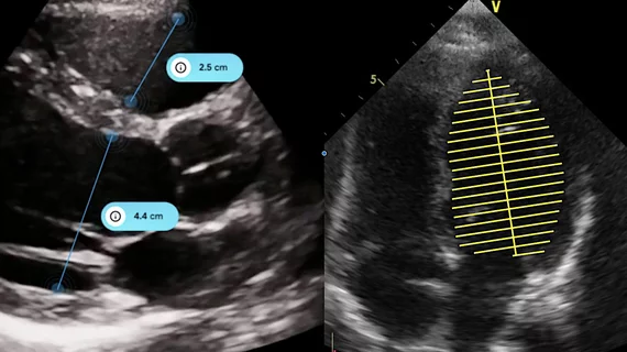
Echocardiography

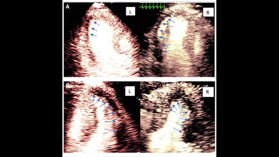
Cardiac ultrasound uses reflected sound waves (echos) to create images of anatomy inside the body. Echocardiograms are the primary cardiac imaging modality used to assess the heart and diagnose or track cardiac issues. Echo is the gold standard imaging modality to assess the heart, particularly with calculating left ventricular ejection fraction (LVEF), which is a measure of cardiac output. In addition to noninvasive standard transthoracic echo (TTE), invasive transesophgeal echo (TEE) is also used when clearer, more detailed imaging of the heart is needed. Both 3D and 4D echo echo systems are rapidly gaining wider adoption and enable new types of assessments, especially in the structural heart space and in transcatheter procedural guidance. Find news on general ultrasound imaging.

Echocardiography: A Beat Ahead in the 21st Century

With better resolution than MR and CT, echocardiography is poised to take the lead in screening and diagnosing heart disease.

Managing Dynamic Images in the Echo Lab

Faster image retrieval, reductions in transcription costs and eliminating repeat studies are among the benefits of ridding your department of videotapes in favor of digitally managing echo exams.

Around the web

The new technology shows early potential to make a significant impact on imaging workflows and patient care. 

Richard Heller III, MD, RSNA board member and senior VP of policy at Radiology Partners, offers an overview of policies in Congress that are directly impacting imaging.
 

The two companies aim to improve patient access to high-quality MRI scans by combining their artificial intelligence capabilities.