Cardiac Imaging

While cardiac ultrasound is the widely used imaging modality for heart assessments, computed tomography (CT), magnetic resonance imaging (MRI) and nuclear imaging are also used and are often complimentary, each offering specific details about the heart other modalities cannot. For this reason the clinical question being asked often determines the imaging test that will be used.

SPECT helpful in staging PCI candidates

SPECT can identify which patients would benefit more from undergoingpercutaneous coronary intervention (PCI) than receiving therapy alonefor blocked coronary arteries, according to late-breaking trial resultspresented at the American Heart Association (AHA) conference inOrlando, Fla., this week.

Cardiac Imaging Goes Straight to the Heart

With heart disease still the leading cause of death in the United States—as well as many other countries—imaging and health IT companies are putting forth an all-out effort to develop and connect cutting-edge diagnostic technology for cardiology. At the forefront of R&D efforts are ways to diagnose cardiovascular disease earlier with more and better information.Vendors are showing great progress as they continue to arm practitioners with newer, better, faster solutions for cardiac care. Among the inroads are enhancements for CT and angiography, upgraded clinical IT systems geared for cardiology and a host of easy-to-use advanced visualization and analysis tools that show more and do more.

Cardiac MRI shows promise for left ventricular noncompaction staging

Left ventricular noncompaction syndrome, a cardiomyopathy characterizedby deep trabeculations in the ventricular wall, is typically diagnosedby echocardiography. However, according to research published thismonth in the American Journal of Roentgenology, echocardiography may not visualize the apical region optimally, leading to underestimation of the degree of the disease. 

Clinicians question utilization of 18F-FDG imaging alone for myocardial viability

Myocardial tissue is morphologically homogenous, yet the heterogeneousuptake of 18F-FDG in the myocardium of healthy subjects is frequentlyobserved. A team of Italian researchers, in a study published thismonth in the Journal of Nuclear Medicine, sought to determineif this impacts the proposed use of 18F-FDG distribution alone for thepurpose of determining myocardial viability.

Mechanical vs. biological approaches in the future of interventional cardiology

Frank Giordano, MD, assistant professor of cardiovascular medicine anddirector of the molecular cardiology program at Yale University Schoolof Medicine, suggested that the future of interventional cardiologywill be a synergism of biology and devices. In a counterpointpresentation, Daniel, Burkhoff, MD, associate professor of medicine atColumbia University in New York City said that device-based therapiesprovide the most realistic methods for the foreseeable future in thefield.  

MRI could set stage for directing unrecognized myocardial infarction treatment

A prospective study investigating the use of MRI to detect clinicallyunrecognized myocardial infarctions has laid the groundwork todetermine if these findings have a different pathogenesis thanrecognized myocardial infarctions.

Vital integrates with Sectra PACS

Sectra has signed an agreement with the Minneapolis-based Vital Imagesunder which Sectra will be able to integrate Vital Images’ Vitreasoftware as part of Sectra PACS.

A case to use MRI for vascular intervention with XFM

For vascular intervention, MRI should be used for diagnoses,particularly with x-ray fiducial markers (XFM), which fuses x-raytechnology with MRI, according to Robert Lederman, MD, clinicalresearch branch of the National Heart and Blood Institute. 

Around the web

The nuclear imaging isotope shortage of molybdenum-99 may be over now that the sidelined reactor is restarting. ASNC's president says PET and new SPECT technologies helped cardiac imaging labs better weather the storm.

CMS has more than doubled the CCTA payment rate from $175 to $357.13. The move, expected to have a significant impact on the utilization of cardiac CT, received immediate praise from imaging specialists.

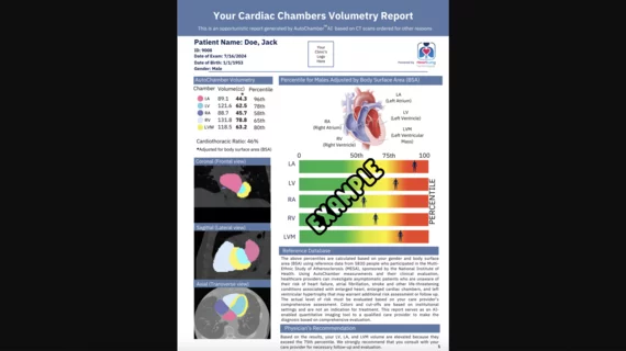
The newly cleared offering, AutoChamber, was designed with opportunistic screening in mind. It can evaluate many different kinds of CT images, including those originally gathered to screen patients for lung cancer. 

Trimed Popup
Trimed Popup