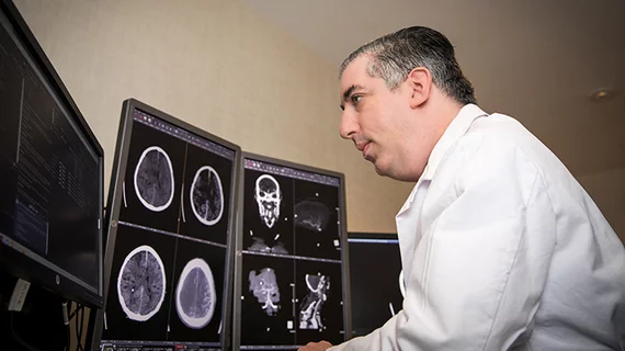
New CT protocol uses scout images to expedite stroke patients' path to MRI
Researchers at the Mayo Clinic have developed a rapid CT protocol to expedite the process of safely getting "wake-up stroke" patients an MRI exam, according to work published this week in Current Problems in Diagnostic Radiology.
Since these specific patients might not be candidates for intravenous thrombolysis, timely diagnosis is critical. MRI is beneficial for identifying a timeline of onset but many patients with symptoms are unable to communicate their MRI risk factors.
“One of the most significant time barriers for emergent MRI is timely screening patients for non-MRI safe implants, which must be excluded to protect the patient from receiving harm from the MRI magnet,” Rahul B. Singh, MBBS, with the Department of Radiology at Mayo Clinic in Jacksonville, and co-authors explained.
When there is no reliable documentation or history clearing a patient for MRI, plain films and CTs must be utilized to rule out metal implantation devices. For these cases, experts at Mayo Clinic developed an MRI-based triage system.
Ideally, within 4.5 hours of symptom onset, patients presenting with signs of an acute stroke first receive a non-contrast head CT to rule out hemorrhage. This is followed by a CTA of the head and neck to monitor any large vessel occlusion.
Perhaps the biggest adjustment to their protocol includes a scout image: any patients presenting for CT scans to rule out stroke also received a quick, full-body CT scan to check for metallic implants.
Once the CT scout image is completed, a radiology resident reviews the exam to clear the patient for MRI. When patients get to the magnet, they are then evaluated for DWI-FLAIR mismatch, which would determine their candidacy for thrombolytic therapy.
“The images can be quickly reviewed to expedite MRI screening,” the doctors wrote. “This is expected to be a significant modification to MRI safety screening protocol in-hospital care that should markedly improve current delays in screening patients.”
You can read the full report in Current Problems in Diagnostic Radiology.
Related Stroke Imaging Content:
Automated CT scoring system accurately predicts prognosis in stroke patients
New CT protocol uses scout images to expedite stroke patients' path to MRI
What decades of data tell us about stroke rates in the U.S.
Most Americans live within an hour of a stroke center
FDA warns providers about potential misuse of imaging-based software for stroke triage
Radiologists must change their approach to stroke care in the AI era
MRI findings associated with poor thrombectomy outcomes after stroke
Risk of recurrent stroke 48% higher among young marijuana users

