Computed Tomography

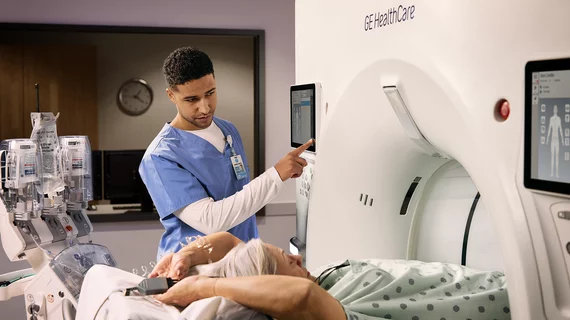
Computed tomography (CT) is a fast and accurate imaging modality often used in emergency settings and trauma imaging. CT scans, with or without (or both) iodinated contrast are frequently used to image the brain, chest, abdomen and pelvis, but also have post-imaging reconstructive capabilities for detailed orthopedic imaging. It is now a standard imaging modality in emergency rooms to quickly assess patients. CT uses a series of X-ray images shot as the gantry rotates around the patient. Computer technology assembles these into into a dataset volume than can be slices on any access, or advanced visualization software can extract specific parts of the anatomy for study. Find more content specific to cardiac CT.
CT scan of a Coca-Cola bottle rectal foreign body. There are a sizable number of emergency room cases each year for items people insert into their rectums that become lodged or lost inside the colon. This includes many types of household items beyond vibrators and commercially made butt plugs. Ghonaim E, Rectal foreign body. Case study, Radiopaedia.org.

PHOTO GALLERY: Abdominal, pelvic and rectal foreign bodies

This is a clinical photo gallery showcasing the uncommon findings of foreign bodies in pelvic and abdominal CT and X-ray, often from emergency room visits. 

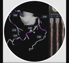
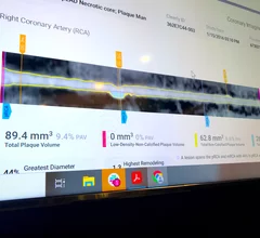
HeartFlow introduced its next generation artificial intelligence (AI) Plaque Analysis with an interactive experience at SCCT 2024. It shows a 3D plaque model and analysis by territory across calcified, non-calcified and low-attenuation plaques. This includes viewing cross-sectional, color-coded images of each plaque type where it was quantified along the vessel. #SCCT #SCCT24 #SCCT2024

Updated HeartFlow technology offers interactive features, full integration with FFR-CT 

HeartFlow is using SCCT 2024 to introduce the world to its updated Plaque Analysis platform. “Accurately diagnosing a patient’s risk for coronary artery disease is critical for determining the best treatment," Chief Medical Officer Campbell Rogers, MD, explained.

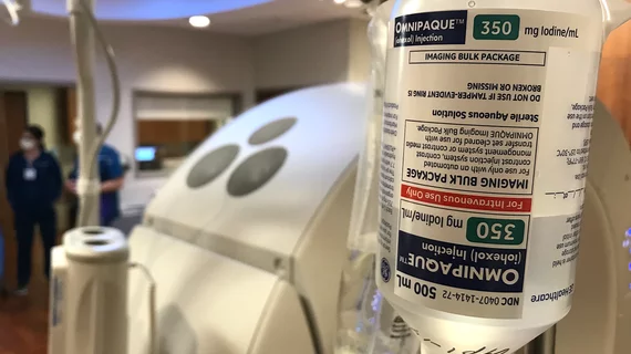
Contrast protocols based on lean body weight save supplies, reduce patient risk

Adjusting contrast dose based on a patient’s lean body weight provides the same image quality as other administration methods, new data show.

Thumbnail

Former smokers who vape now still need to be screened for lung cancer. Many of them aren't

Despite kicking their initial habit, these individuals remain at risk of developing lung cancer due to their history of smoking cigarettes, current use of e-cigarettes, or both, experts caution.

Thumbnail

New study calls ED imaging protocols for geriatric head injuries into question

The new data suggest that admitting patients for observation and additional imaging to monitor for delayed intracranial hemorrhage might often be unnecessary.  

The feasibility of reduced contrast flow rates in PE studies

Around one out of every 1,000 people in the U.S. are diagnosed with a pulmonary embolism every year, but even more undergo imaging to rule out clots.  

Imaging study reveals 'significant deformities' in lungs of some COVID patients

These changes could have long-lasting implications for lung function, authors of the new study warn.

EHR interventions increase lung cancer screening by 30% but still leave over half of patients behind

Although CT lung cancer screening is known to improve detection rates and health outcomes, compliance among eligible patients remains lackluster.

Around the web

RBMA President Peter Moffatt discusses declining reimbursement rates, recruiting challenges and the role of artificial intelligence in transforming the industry.

Deepak Bhatt, MD, director of the Mount Sinai Fuster Heart Hospital and principal investigator of the TRANSFORM trial, explains an emerging technique for cardiac screening: combining coronary CT angiography with artificial intelligence for plaque analysis to create an approach similar to mammography.

A total of 16 cardiology practices from 12 states settled with the DOJ to resolve allegations they overbilled Medicare for imaging agents used to diagnose cardiovascular disease.