Computed Tomography

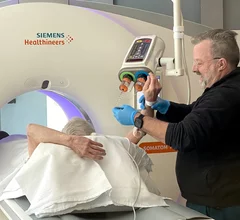
Computed tomography (CT) is a fast and accurate imaging modality often used in emergency settings and trauma imaging. CT scans, with or without (or both) iodinated contrast are frequently used to image the brain, chest, abdomen and pelvis, but also have post-imaging reconstructive capabilities for detailed orthopedic imaging. It is now a standard imaging modality in emergency rooms to quickly assess patients. CT uses a series of X-ray images shot as the gantry rotates around the patient. Computer technology assembles these into into a dataset volume than can be slices on any access, or advanced visualization software can extract specific parts of the anatomy for study. Find more content specific to cardiac CT.
Thumbnail

AI model slashes cerebral aneurysm interpretation times

Similar tools could have great utility in enhancing clinical workflows and improving the diagnosis of cerebral aneurysms, which can be deadly if ruptured.

Radiomics-based tool predicts fracture risk in COPD patients

Such technology can be leveraged to improve risk assessments without accruing additional expenses for patients or clinicians, potentially filling in gaps where DXA exams fall short.

Example of an automated artificial intelligence (AI) assessment of soft coronary plaque from a CT scan from the vendor Cleerly. The AI gives a very detailed report of all the plaque in all the coronary vessels. Some cardiology experts believe this may be the way of the future in screening patients for early coronary disease and monitoring the impact of prevention efforts such as statins to determine if more aggressive treatments are needed.

Medicare administrative contractors approve coverage of AI-enabled quantitative CT

Four of the seven Medicare Administrative Contractors (MACs) announced they will now cover artificial intelligence-enabled quantitative coronary tomography (AI-QCT) and coronary plaque analysis (AI-CPA). 

Ron Blankstein, MD, Brigham and Womens Hospital, explains a study using AI opportunistic screening in non-cardiac CT scans looking for coronary artery disease.

Use of AI opportunistic screening in CT for cardiovascular disease

Ron Blankstein, MD, professor of radiology, Harvard Medical School, explains the use of artificial intelligence to detect heart disease in non-cardiac CT exams.

Banner ASC in Sun City, Arizona.

Low doses of radiation still increase risk of leukemia, lymphoma and myeloma

A new study found cumulative radiation exposure of 16 mSv to red bone marrow can cause an increased rate of some hematological malignancies.

Thumbnail

Incidental finding characteristics that warrant communication

Three factors are associated with true high-risk findings—imaging protocols for aortic valve implantation planning, imaging area, and cardiology department orders.

Left, coronary CT angiography of a vessel showing plaque heavy calcium burden. Right, image showing color code of various types of plaque morphology showing the complexity of these lesions. The right image was processed using the FDA cleared, AI-enabled plaque assessment from Elucid.

FDA clears new software for AI-powered CCTA assessments

Elucid's PlaqueIQ was trained to turn CCTA images into interactive 3D reports that help physicians visualize the presence of atherosclerosis.

Deep learning denoising produces quality trauma head CT images at 25% of the radiation dose

Denoising algorithm produces quality trauma head CTs at a fraction of the radiation dose

Radiation doses needed for quality head CT scans in trauma settings can be reduced by as much as 75% using DL-based denoising.

Around the web

The nuclear imaging isotope shortage of molybdenum-99 may be over now that the sidelined reactor is restarting. ASNC's president says PET and new SPECT technologies helped cardiac imaging labs better weather the storm.

CMS has more than doubled the CCTA payment rate from $175 to $357.13. The move, expected to have a significant impact on the utilization of cardiac CT, received immediate praise from imaging specialists.

The newly cleared offering, AutoChamber, was designed with opportunistic screening in mind. It can evaluate many different kinds of CT images, including those originally gathered to screen patients for lung cancer. 

Trimed Popup
Trimed Popup