Computed Tomography

Computed tomography (CT) is a fast and accurate imaging modality often used in emergency settings and trauma imaging. CT scans, with or without (or both) iodinated contrast are frequently used to image the brain, chest, abdomen and pelvis, but also have post-imaging reconstructive capabilities for detailed orthopedic imaging. It is now a standard imaging modality in emergency rooms to quickly assess patients. CT uses a series of X-ray images shot as the gantry rotates around the patient. Computer technology assembles these into into a dataset volume than can be slices on any access, or advanced visualization software can extract specific parts of the anatomy for study. Find more content specific to cardiac CT.
Thumbnail

Routine abdominal CTs offer insight into cardiovascular risk

During this year's RSNA meeting, providers were especially interested in how AI applications could enhance opportunistic screening capabilities.

New scoring system accurately predicts coronary risk in women

The system combines findings from SPECT myocardial perfusion imaging with lifestyle and clinical factors to predict risk of major cardiovascular events.

Silicosis on CT

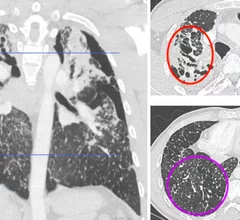
Abnormal CT findings increasingly common among countertop workers

Individuals who work with engineered stone countertops are increasingly being diagnosed with silicosis—a chronic lung condition that is the result of inhaling large amounts of silica dust.

Philips CT 5300

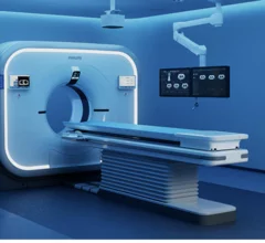
New AI-enabled CT system is officially available in North America

The scanner is equipped with features aimed at addressing some of the biggest pain points of both radiologists and technologists.

Incidental interstitial lung abnormalities largely underreported

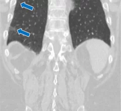
Less than half of incidental interstitial lung abnormalities on CT exams are reported

This is despite experts cautioning that these findings should be "systematically and fully assessed" due to their progressive nature.

More than 1/3 of old strokes are incidentally detected on head CT

A new analysis in JACR estimates that spotting the signs of a previously undetected stroke could benefit up to 200,000 people in the United States every year.

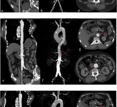
Deep learning recons reduce contrast and radiation doses in aortic CTA

Deep learning reconstruction cuts radiation and contrast dose by half in aortic CTA exams

A new analysis in Academic Radiology dives into the importance of finding balance between reducing radiation exposure while maintaining image quality. 

Dual energy CT parameters help determine the type of clot behind strokes

Knowledge of clot composition helps guide clinical management decisions following acute ischemic stroke.

Around the web

GE HealthCare designed the new-look Revolution Vibe CT scanner to help hospitals and health systems embrace CCTA and improve overall efficiency.

Clinicians have been using HeartSee to diagnose and treat coronary artery disease since the technology first debuted back in 2018. These latest updates, set to roll out to existing users, are designed to improve diagnostic performance and user access.

The cardiac technologies clinicians use for CVD evaluations have changed significantly in recent years, according to a new analysis of CMS data. While some modalities are on the rise, others are being utilized much less than ever before.