Computed Tomography

Computed tomography (CT) is a fast and accurate imaging modality often used in emergency settings and trauma imaging. CT scans, with or without (or both) iodinated contrast are frequently used to image the brain, chest, abdomen and pelvis, but also have post-imaging reconstructive capabilities for detailed orthopedic imaging. It is now a standard imaging modality in emergency rooms to quickly assess patients. CT uses a series of X-ray images shot as the gantry rotates around the patient. Computer technology assembles these into into a dataset volume than can be slices on any access, or advanced visualization software can extract specific parts of the anatomy for study. Find more content specific to cardiac CT.
Thumbnail

New study calls ED imaging protocols for geriatric head injuries into question

The new data suggest that admitting patients for observation and additional imaging to monitor for delayed intracranial hemorrhage might often be unnecessary.  

The feasibility of reduced contrast flow rates in PE studies

Around one out of every 1,000 people in the U.S. are diagnosed with a pulmonary embolism every year, but even more undergo imaging to rule out clots.  

Imaging study reveals 'significant deformities' in lungs of some COVID patients

These changes could have long-lasting implications for lung function, authors of the new study warn.

EHR interventions increase lung cancer screening by 30% but still leave over half of patients behind

Although CT lung cancer screening is known to improve detection rates and health outcomes, compliance among eligible patients remains lackluster.

Older adults among the walking wounded with incident TBI

The demographic most at risk seems to be White women who are healthy, active and of high socioeconomic status.

Thumbnail

Wake Forest receives $1.5 million to fund imaging study aimed at bone loss after bariatric surgery

The funding will go toward the Strategies to Reduce the Onset of Sleeve Gastrectomy Associated Bone Loss (STRONG BONES) trial.

Lung cancer screening CT image in a 66-year-old male patient shows a sessile nodule with internal air in the left mainstem to left upper lobe bronchus (arrow) with a mean diameter of 10 mm. The nodule was assigned as Lung-RADS category 4A in the clinical report. (B) Follow-up CT image shows the lesion is resolved. Image courtesy of RSNA

Lung-RADS update helps limit false-positive results, unnecessary procedures

Using the latest version was associated with improved diagnostic accuracy, researchers wrote in a new analysis. 

Dual energy CT scanners should be powered down more often to reduce energy consumption.

Why some CT equipment should be powered down more often

And not just on nights and weekends, authors of a new analysis on energy consumption in CT departments argue.

Around the web

The nuclear imaging isotope shortage of molybdenum-99 may be over now that the sidelined reactor is restarting. ASNC's president says PET and new SPECT technologies helped cardiac imaging labs better weather the storm.

CMS has more than doubled the CCTA payment rate from $175 to $357.13. The move, expected to have a significant impact on the utilization of cardiac CT, received immediate praise from imaging specialists.

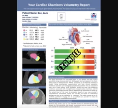
The newly cleared offering, AutoChamber, was designed with opportunistic screening in mind. It can evaluate many different kinds of CT images, including those originally gathered to screen patients for lung cancer. 

Trimed Popup
Trimed Popup