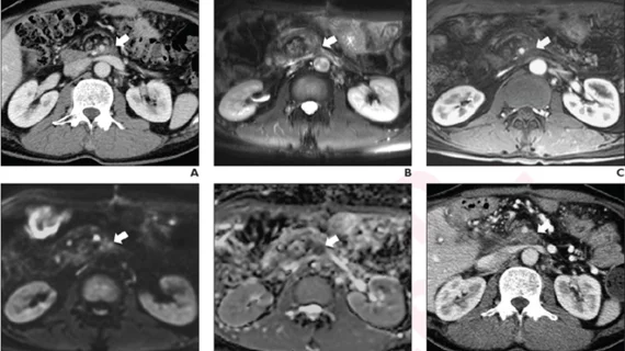
Diffusion-weighted MRI boosts detection of locally recurrent pancreatic cancer
For pancreatic cancer patients who have undergone pancreatic ductal adenocarcinoma (PDAC) resection, traditional MRI images alone can present difficulties in distinguishing recurrent tumors from post-surgical fibrosis. A new study published in American Journal of Roentgenology (AJR) found that adding diffusion-weighted MRI (DWI) to traditional MRI may help make that differentiation.[1]
“The findings indicate a potential role for MRI with DWI in surveillance protocols after PDAC resection,” corresponding author Tae Wook Kang, MD, of Samsung Medical Center in Seoul, South Korea, said in a statement about the study’s findings.
To make that determination, Kang and coauthors performed a retrospective study examining 66 patients who had PDAC resection between January 2009 and March 2016, and for whom a postoperative surveillance CT revealed a soft tissue lesion at the operative site or at the site of peripancreatic vessels. Those patients were then referred for MRI with DWI for additional evaluation.
Two observers independently reviewed the conventional MRI images versus the MRI with DWI images, using CT at least six months after MRI as a reference standard.The MRI with DWI images showed higher sensitivity, without difference in specificity, in detecting local recurrence after PDAC resection, the study concluded.
Based on those results, using MRI with DWI may be able to play an important role in helping providers identify recurrent pancreatic cancer at an earlier stage.
“MRI with DWI as a problem-solving tool during post-operative surveillance after PDAC resection could facilitate earlier detection of recurrences,” the study’s authors added, “guiding prognostic assessment and treatment decisions.”
Reference:
1. Shin, Nari; Kang, Tae Wook; Min, Ji Hye; et al. Utility of Diffusion-Weighted MRI for Detection of Locally Recurrent Pancreatic Cancer After Surgical Resection. American Journal of Roentgenology. May 25, 2022. DOI: 10.2214/AJR.22.27739
Related Pancreatic Cancer Content:
AI spots pancreatic cancer in its earliest stages
Delayed-phase contrast-CT scans can help detect early pancreatic cancers, study shows
CT imaging features forecast outlook for patients with aggressive pancreatic cancer
MR imaging-guided SABR extends the lives of patients with inoperable pancreatic cancer
Radiology reporting template bolsters follow-up care for incidental pancreatic findings
