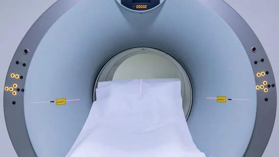
Improving MRI reconstructions by combining traditional techniques with new ML tools
Researchers are working on new ways to use machine learning techniques to make MRI exams faster by improving upon current reconstruction methods.
The details were shared recently in the Proceedings of the National Academy of Sciences of the United States of America (PNAS). In the paper, researchers discussed the sequential manner in which MRI scans acquire data, which in turn increases acquisition times. With machine learning techniques, experts can overcome this time burden by essentially filling in the blanks during image reconstruction, rather than capturing every frequency during the scan. Both methods are intended to achieve the same goal, yet neither comes without flaws.
Traditionally, experts have used compressed sensing methods to make MRI acquisitions faster. Simply put, this method makes images smaller. In contrast, machine learning techniques are capable of bypassing frequencies during MRI exams by utilizing algorithms trained to predict results from in between the skipped frequencies.
While deep learning techniques have been shown to be superior to compressed sensing, many experts in the field have voiced concerns over potential bias that could arise depending on the data used to train the algorithm. This would result in the algorithm misinterpreting images.
Meeting In The Middle
Researchers with the University of Minnesota Twin Cities have been working to fine-tune a method that includes both traditional reconstruction techniques with advancements in data tools and machine learning. The newer technique uses linear representations, a small number of parameters and enables an explainable convex optimization procedure at inference time. Experts involved in the research shared that the results have thus far been positive to accelerating MRI exams without sacrificing quality.
“We found that if you tune the classical methods, they can perform very well,” explained the paper’s senior author, Mehmet Akcakaya, the Jim and Sara Anderson Associate Professor in the University of Minnesota Department of Electrical and Computer Engineering. “So, maybe we should go back and look at the classical methods and see if we can get better results. There is a lot of great research surrounding deep learning as well, but we’re trying to look at both sides of the picture to see where we can find the best performance, theoretical guarantees, and stability.”
The detailed research is available here.

