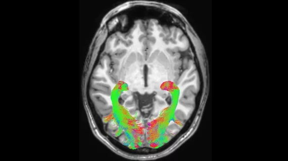
New 7T MRI scanners could increase radiologists' role in treating neurological conditions
The recent emergence of powerful scanners, such as 7T MRIs, could play a pivotal role in developing treatments for Parkinson’s disease and similar conditions.
New research published in Movement Disorders utilized 7T MRI brain scans to analyze the locus coeruleus of patients with Parkinson’s and progressive supranuclear palsy (PSP). The locus coeruleus is located in the back of the brain and is very small, making it difficult to accurately visualize using 3T scanners. This part of the brain is of particular interest to researchers because it produces noradrenaline, which plays a role in attention, arousal, thinking, motivation and other brain functions that tend to diminish in patients with Parkinson’s and PSP.
“The locus coeruleus is a devil to see on a normal scanner,” said Professor James Rowe from the Department of Clinical Neurosciences at the University of Cambridge, who led the study. “Even good hospital scanners just can't see it very well. And if you can't measure it, you can't work out how two people differ: who's got more, who's got less? We’ve wanted MRI scanners to be good enough to do this for some time.”
Previous research conducted by Professor Rowe examined donated brains of people who had been diagnosed with PSP and found that the locus coeruleus (LC) had significantly diminished in some of the patients. The issue that Rowe and his colleagues faced was analyzing that region of the brain in living patients, as most MRI scanners did not provide appropriate resolution. This is where newer, more powerful 7T MRI machines come into play.
Using a 7T scanner, researchers completed scans on the brains of 25 people with idiopathic PD, 14 with probable PSP and 24 age-matched healthy controls. The participants also underwent clinical and cognitive assessments.
The imaging revealed significant LC degeneration in the caudal subregion in both the PD and PSP individuals compared to controls. These findings were found to be strongly associated with worse cognition and higher apathy scores.
The authors explained that previous research pertaining to nonandrogenic treatments in patients with similar symptoms to those seen in patients with LC damage have proven to be safe and effective. The ability to better visualize the LC in patients with PD and PSP has enabled the scientists to continue their research efforts by identifying candidates who may benefit from similar noradrenergic therapeutic strategies. This theory is being tested by Rowe and colleagues in another clinical trial at Cambridge University Hospitals NHS Foundation Trust.
Corresponding author Rong Ye, PhD, from the Department of Clinical Neurosciences and Cambridge University Hospitals NHS Trust, University of Cambridge, spoke of the machine’s benefits, stating, “the ultra-powerful 7T scanner may help us identify those patients who we think will benefit the most. This will be important for the success of the clinical trial, and, if the drugs are effective, will mean we know which patients to give the treatment to.”
Related neuroimaging news:
Veterans who experience chronic pain and trauma have connectivity abnormalities on brain imaging
New MRI technique helps physicians ID multiple sclerosis lesions
Memory complaints associated with structural brain abnormalities and increased dementia risk
References:
Ye, R & O’Callaghan, C et al. Locus Coeruleus Integrity from 7T MRI Relates to Apathy and Cognition in Parkinsonian Disorders. Movement Disorders; 17 May 2022; DOI: 10.1002/MDS.29072

