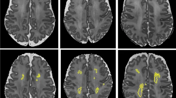
New MRI technique earlier detects multiple sclerosis, potentially improving treatment approaches
Researchers in Austria have unveiled an exciting new MRI technique capable of detecting early biochemical changes in the brains of people diagnosed with multiple sclerosis, which could lead to faster, more targeted treatments.
The research, published Tuesday in Radiology, was conducted around the premise of MR spectroscopic imaging (MRSI), and its ability to look beyond the white and gray matter of the brain in order to detect and evaluate metabolites. For this particular study, researchers used a 7-Tesla (T) magnet.
"MRI of neurochemicals enables the detection of changes in the brain of multiple sclerosis patients in regions that appear inconspicuous on conventional MRI," explained senior author Wolfgang Bogner, PhD, from the High Field MR Centre at the Medical University of Vienna.
A total of 65 patients with MS and 20 control patients were included in the study. The goal was to evaluate any pathologic disturbances in the normal-appearing white matter (NAWM) and cortical gray matter (CGM). and connect any dots that could link those changes to the severity of a patient’s disability.
In patients with MS, higher levels of myo-inositol (mI) were observed in the NAWM and the team reported an increased ratio of ml to creatinine. At the same time, lower levels of N-acetylaspartate (NAA) were noted, which is known to affect the integrity of neurons in the brain. These changes did appear to correlate with patient disability.
"Some neurochemical changes, particularly those associated with neuroinflammation, occur early in the course of the disease and may not only be correlated with disability, but also be predictive of further progression such as the formation of multiple sclerosis lesions," lead author Eva Heckova, PhD, from the High Field MR Centre at the Medical University of Vienna, said of the new technique.
Typically, it is focal white matter lesions on brain imaging that doctors often see as an indication of MS, but those do not fully align with disease severity. This new imaging technique, however, gives clinicians a more in-depth understanding of the pathologic manifestations of MS. It could allow them to detect the disease earlier and fine tune their treatment plans, the authors suggested.
“We will continue our ongoing longitudinal clinical study to validate its ability to detect clinically important pathologic changes in the brain of multiple sclerosis patients and to evaluate the efficacy of different treatment regimens earlier than other currently available clinical tools," Heckova said.
You can view the detailed research in Radiology.

