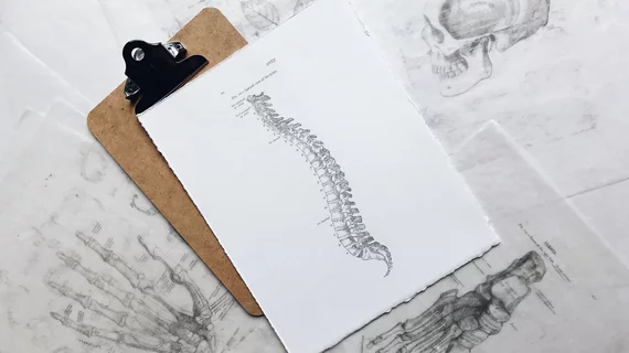
Conventional radiography superior to MRI for certain spinal imaging, new research shows
Imaging morphologic changes of the spine can be achieved through a number of modalities but recent research suggests that a combination of conventional radiography and magnetic resonance imaging may be best.
A study published in Insights Into Imaging compared the reliability of conventional radiography versus MRI when visualizing vertebral lesions in patients with axial spondyloarthritis (SpA) and did not find one to be superior to the other. Instead, researchers suggest using routine X-ray imaging in conjunction with MRI.
“MRI has the potential to detect bone marrow edema and osseous changes such as fatty lesions, erosions and sclerosis prior to their manifestation on conventional X-rays, while avoiding radiation exposure,” corresponding author, Michael Fuchsjäger, with the Department of Radiology at Medical University of Graz, and co-authors explained. “However, conventional radiography seems to better depict structural damage, such as syndesmophytes, because of better spatial resolution than MRI.”
The study included 40 patients who had been diagnosed with SpA. Conventional X-rays of the cervical and lumbar spine were obtained for every patient, followed by an MRI. Exams were analyzed by two radiologists.
The most common X-ray finding was the presence of lumbar syndesmophytes, followed by cervical syndesmophytes. MRI exams better visualized thoracic syndesmophytes that are not typically seen on conventional radiographs.
Inflammatory changes were better depicted on MRI, while miniscule bony changes were better visualized on conventional X-ray.
“In the cervical and lumbar spine, conventional radiography seems superior to MRI in depicting morphologic changes, whereas MRI may visualize highly prevalent morphologic changes in the thoracic spine in addition to inflammatory vertebral lesions,” the experts explained.
The earlier these developments are discovered the better patients fare long term. Using both conventional X-ray and MRI as complementary diagnostic tools could help to detect morphologic changes earlier and improve overall patient prognosis, the authors noted.

