Molecular Imaging

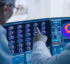
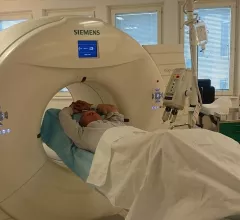
Molecular imaging (also called nuclear medicine or nuclear imaging) can image the function of cells inside the body at the molecular level. This includes the imaging modalities of positron emission computed tomography (PET) and single photon emission computed tomography (SPECT) imaging. How does PET and SPECT imaging work? Small amounts of radioactive material (radiopharmaceuticals) injected into a patient. These can use sugars or chemical traits to bond to specific cells. The radioactive material is taken up by cells that consume the sugars. The radiation emitted from inside the body is detected by photon detectors outside the body. Computers take the data to assemble images of the radiation emissions. Nuclear images may appear fuzzy or ghostly rather than the sharper resolution from MRI and CT.  But, it provides metabolic information at a cellular level, showing if there are defects in the function of the heart, areas of very high metabolic activity associated with cancer cells, or areas of inflammation, data not available from other modalities. These noninvasive imaging exams are used to diagnose cancer, heart disease, Alzheimer’s and Parkinson’s disease, bone disorders and other disorders. 

ITM to supply Alpha-9 radioisotopes to develop new cancer drugs

The German manufacturer and Canadian drug developer are partnering to address the supply crunch of radioisotopes.

Isotope used in cancer drug development shipped for clinical trials

Actinium-225 remains in short supply, slowing drug research.

Bhvita Jani, medical imaging principal analyst, Signify Research, discusses growing trend of theragnostics in nuclear imaging and oncology at RSNA2023. #theragnostics #RSNA #RSNA23 #RSNA2023 #radiology #NucMed #Oncology

Theragnostics: A growing trend in molecular imaging

Bhvita Jani, medical imaging principal analyst at Signify Research, discusses the evolution of theragnostics and its applications in nuclear imaging and oncology.

Thumbnail

SPECT/CT helps personalize treatment options for prostate cancer patients

Using SPECT/CT imaging, providers can stratify patients based on their treatment responses and make personalized adjustments accordingly.

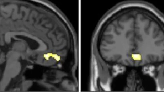
molecular imaging study on brain connections in obese individuals

Obesity linked to altered brain connectivity on PET/MRI exams

New research, presented at SNMMI 2023, details how the brains of obese individuals differ from those who maintain a healthy weight.  

SNMMI Image of the Year

SNMMI unveils Image of the Year

This year the Henry N. Wagner Jr., Image of the Year is actually a group of images displaying the efficacy of a 68Ga-DOTA-5G and 177Lu-DOTA-ABM-5G theranostic pairing that improves the detection and treatment of metastatic pancreatic cancer. 

whole-body pet scanner add-on device presented at #SNMMI 2023

New PET technology offers 'clear improvement' of whole-body scan resolution

The new technology achieves this by way of two add-on detectors that simultaneously scan patients during their whole-body PET exam.

Thumbnail

New prostate cancer PET imaging agent officially available for commercial use

Posluma (flotufolastat F 18) received the U.S. Food and Drug Administration’s approval in May 2023.

Around the web

The nuclear imaging isotope shortage of molybdenum-99 may be over now that the sidelined reactor is restarting. ASNC's president says PET and new SPECT technologies helped cardiac imaging labs better weather the storm.

CMS has more than doubled the CCTA payment rate from $175 to $357.13. The move, expected to have a significant impact on the utilization of cardiac CT, received immediate praise from imaging specialists.

The newly cleared offering, AutoChamber, was designed with opportunistic screening in mind. It can evaluate many different kinds of CT images, including those originally gathered to screen patients for lung cancer. 

Trimed Popup
Trimed Popup