Oncology Imaging

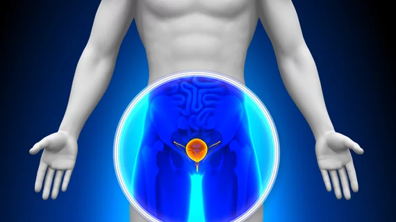
Medical imaging has become integral to cancer care, assessing the stage and location of cancerous tumors. By utilizing powerful imaging modalities including CT, MRI, MRA and PET/CT, oncology imaging radiologists are able to assist referring physicians in the detection and diagnosis of cancer.
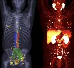
Thumbnail

Prostate cancer size is frequently underestimated on imaging

Newly published data recently revealed just how common the extent of prostate cancer is underestimated based on imaging measurements. 

Two examples of PSMA-PET scans showing numerous prostate cancer metastases spread throughout the body. Many of these smaller tumors would not have been dected on previous standard-of-care imaging. Photo on left courtesy of SNMMI, right University of Chicago. #PSMAPET

PSMA-PET a more cost-effective option for patients long-term compared to standard prostate imaging

The findings support adopting F-18 DCFPyL PET/CT as the standard of care for prostate cancer staging, authors of a new Scientific Reports paper concluded.

Thumbnail

Accessibility, reimbursement and other issues limit uptake of PET/CT among oncologists, survey reveals

Difficulty obtaining PET/CT scans was reported by 55% of respondents, with 21% citing this as the greatest barrier in treating classic Hodgkin lymphoma.

shear-wave elastography TIRADS

Shear wave elastography assessment improves TI-RADS scoring

That’s according to an award-winning scientific online poster presented this week during the American Roentgen Ray Society’s annual meeting being held in Honolulu, Hawaii.

lung cancer imaging features associated with worse outcomes following missed diagnosis

Some imaging features of missed lung cancer indicate worse outcomes

Features pertaining to location, density and superimposed structures were recently found to be associated with poorer outcomes for patients who initially had their lung cancer overlooked on radiographs.

Thumbnail

Experts release new appropriate use criteria for lymphoscintigraphy in sentinel node mapping

The AUC includes a total of 32 clinical scenarios that have been grouped into four categories of cancer: breast, skin, cancer of other sites and lymphedema.

x-ray dosimetry capsule monitors radiation dose during radiotherapy

Swallowable capsule monitors radiation dose in real-time during radiotherapy

Coming in at a diameter of 5 mm and thickness of 0.2 mm, the X-ray dosimeter measures radiation dose up to five times more accurately than standard methods. 

Thumbnail

Imaging considerations included in tattoo guidelines for at-risk cancer population

Cases of mistaken malignancy have become more common as the popularity of tattoos has grown in recent years.

Around the web

The use of advanced AI software to assess CCTA images continues to gain more momentum.

The new guidelines detail the use of echocardiography to evaluate patients for a variety of conditions.

One of the most formidable societies of medical professionals in the U.S. is going toe-to-toe with Robert F. Kennedy’s HHS over changing vaccination recommendations.