
BabyNet AI model predicts fetal birth weight
A group of Dutch researchers from the University of Amsterdam may have devised a method to predict fetal birth weight using ultrasound scans.
Utilizing clinical data to enhance the analysis of ultrasound videos via artificial intelligence, the algorithm was found to be as effective as an experienced physician in predicting details on birth weight. The results are published in Computers in Biology and Medicine. [1]
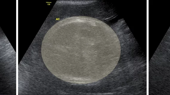
According to the researchers, their method could be effective in assisting physicians with selecting the safest delivery method for mothers. While ultrasound is the typical method for assessing fetal size, it’s challenged by a need to properly measure much of the unborn’s body—such as femur length, head circumference, abdominal circumference, and more—that often require an experienced operator to make the scans, in addition to an expert clinician to properly read them.
“We presented, for the first time, an automated fetal birth weight prediction method that utilizes multimodal data, along with the visual data processing component of our network, which is called BabyNet,” wrote the research team led by Szymon Płotka from the University of Amsterdam, adding that is a “hybrid model that efficiently combines transformers and convolutional neural networks,” two deep-learning methods that are often used to analyze images.
For their study, Płotka and his team used fetal ultrasound video scans conducted within 24 hours of a mother giving birth, inputting the actual birth weight of babies to determine the accuracy of the new BabyNet model versus other algorithms.
BabyNet was given access to clinical data surrounding the fetal size of 194 births, along with 582 ultrasound videos. The analysis of BabyNet was compared to the weight estimated by trained clinicians from four different medical centers, as well as the numbers given by 6 other algorithms that are commonly used to provide fetal birth weight predictions.
The researchers found that BabyNet had significantly higher performance than clinicians in 3 out of 4 of the medical centers (p = 0.09), additionally outperforming all of the other algorithms.
“The use of video analysis in fetal US examinations offered several advantages over static images, one of which was the ability to utilize 2D+t spatio-temporal feature representations that could enhance performance,” the authors wrote, noting that changes in position of the fetus within the 24-hour window complicated the analysis of the BabyNet, which could account for the one comparative scenario where it was outperformed by clinicians.
“While our current work may have some limitations in terms of explainability, we plan to address them in future work to ensure a more transparent and understandable outcome,” the study authors wrote.
The full results can be found here.

Chad is an award-winning writer and editor with over 15 years of experience working in media. He has a decade-long professional background in healthcare, working as a writer and in public relations.