
Why the way microcalcifications on mammograms are regarded could change
New research on the biomineralization of microcalcifications could change how radiologists report the mammographic findings in the future.
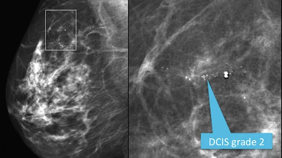
According to experts involved in the new research, published in Science Advances on Feb. 22, the tissue environment where microcalcifications of the breast are formed could hold clues into how breast cancer progresses. Microcalcifications on mammograms typically do not signal malignancy unless they are presented in a specific pattern, but if a more detailed analysis of their origin suggests that the findings could be indicative of cancer risk, that could change how they are regarded in the future, the team suggests.
“Usually after the initial mammogram, microcalcifications are largely ignored. And what we’re saying is we can look beyond the resolution of the mammogram, at the microscopic and chemical level, and get more information from these microcalcifications,” said co-senior author Lara Estroff, professor of materials science and engineering in Cornell Engineering.
The research team specializes in biomineralization, studying how mineral deposits grow within a living organism. Estroff has been collaborating with the paper’s co-senior author Claudia Fischbach, the Stanley Bryer 1946 Professor of Biomedical Engineering, for more than a decade in the study of how breast cancer spreads into bones. During their research, the two discovered bone-like material at primary tumor sites, which prompted them to study these microcalcifications and their surrounding tissue environments further.
This led them to their most recent work studying tissue samples containing microcalcifications from women with breast cancer.
“We said, 'can we take everything that we know from studying physiological biominerals, and apply it now to these pathological minerals?'” Estroff said.
It took many years, many imaging and histology analyses and collaborating with many doctors, including Dr. Daniel Sudilovsky, then at Cayuga Medical Center, to characterize the pathology of the microcalcifications of the tissue samples.
This led to a number of revelations, listed below:
- Microcalcifications associated with cancer tend to cluster into physiological groups that reflect tissue type and local malignancy.
- A variety of mineral carbonates are found in tumors.
- Zinc, iron and aluminum are enhanced in malignant-localized calcifications.
- Worse prognosis is associated with patients who have lower ratios of lipids to proteins in their microcalcifications.
It is not yet known whether the microcalcifications are a result of cancer, or a precursor to it, but the team suggests that their findings indicate there is an association between microcalcifications and disease severity.
It remains to be seen how this will affect radiologists’ reporting of microcalcifications in the future.
To learn more, click here.

In addition to her background in journalism, Hannah also has patient-facing experience in clinical settings, having spent more than 12 years working as a registered rad tech. She began covering the medical imaging industry for Innovate Healthcare in 2021.