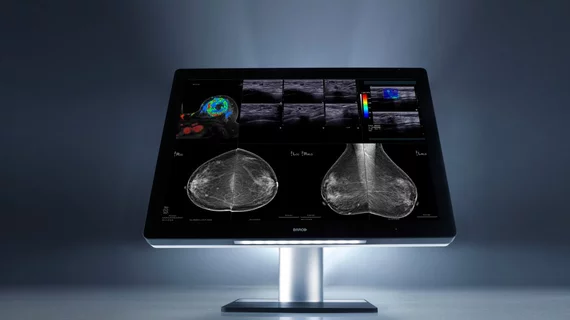
Radiomics differentiates luminal A breast cancer, benign lesions from MRI dataset
Researchers are always looking to improve their diagnoses—and machine learning has a lot of potential to help, alongside radiomic signatures, to aid in diagnosis and prognosis of breast cancers.
Researchers have found that quantitative radiomics can better distinguish between benign lesions and luminal A breast cancers than using maximum linear size alone, according to a study published May 10 in Academic Radiology.
The team of researchers, led by Heather Whitney, PhD, an associate professor of physics at Wheaton College in Wheaton, Illinois, extracted multiple radiomic features from a large breast MRI database for their study.
Ultimately, the team determined that the radiomic feature of irregularity plays an important role in feature selection and computer-aided diagnosis.
"We evaluated the classification of a clinical dataset of lesions as benign versus luminal A using three variations of radiomic signatures: using maximum linear size alone, using feature selection from a full set of radiomic features and using feature selection from radiomic features excluding those describing size," Whitney et al. wrote.
The team collected 212 benign lesions and 296 luminal A breast cancers. They were appropriately grouped from breast MR images, in which 38 radiomic features were extracted. The researchers then performed 10-fold cross validation to distinguish between breast lesions and cancers using radiomics compared with maximum linear size alone.
"Area under the receiver operating characteristics curve (AUC) was used as the figure of merit," the researchers wrote.
The researchers found the following:
- For maximum linear size alone, AUC and 95 percent confidence interval were 0.797, compared to 0.846 and 0.848 for lesion signature feature selection protocols including and excluding size features.
- The irregularity feature was chosen in nine of 10 folds and in all folds when size features were included and excluded.
- AUC for the radiomic signature using feature selection from all features was statistically equivalent to using feature selection from all features excluding size features, within an equivalence margin of 2 percent.

