Triplet pregnancy fetus in fetu: A rare case report
Recently in Cureus, experts described their experience with a case of fetus in fetu (FIF)—a rare occurrence in itself, but this particular case drew added attention because it involved a triplet pregnancy.
This case report occurred in Bahrain [1]. To the clinicians’ knowledge, this was the first case of FIF to be reported within the country. While FIF is an extremely rare condition—occurring in about one in 500,000 births—it is even more rare in triplet pregnancies. Nonetheless, the condition recently presented in a 4-month-old female who was a product of an in vitro fertilization (IVF) twin pregnancy to a 30-year-old primigravida mother.
“Our case is unique because this condition appeared as a part of a triplet pregnancy, which resulted in two viable babies and the third baby was found encased in the body of the largest living newborn,” shared the report’s co-authors Bayan Hasan, MD, with Kims Hospital, and Mohamed Ebrahim, of the Radiology Department at Salmaniya Medical Complex, both in Manama, Bahrain.
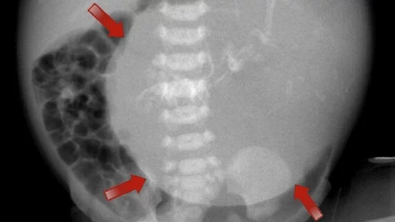
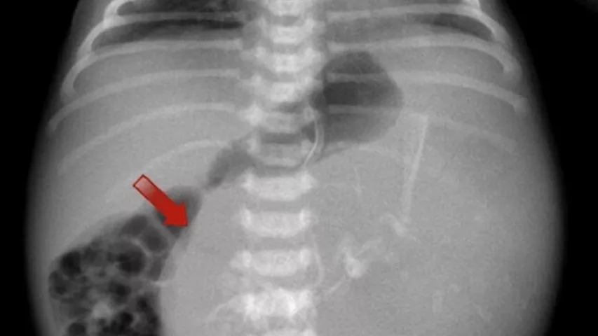
During the patient’s pregnancy, an antenatal abdominal ultrasound revealed an abdominal cystic lesion with ascites in one fetus. Upon the twin of interest’s birth, clinicians noticed that the baby’s abdomen was visibly distended, despite sufficient APGAR scores and an otherwise normal appearance. Further analysis included an abdominal radiograph, which demonstrated a large cystic shadow in the left hypochondrium with radio-opaque shadows.
CT and MRI exams of the abdomen and pelvis showed fat, fluid, soft tissues, and parts of the fetal skeleton and neural tissues within the large mass, which had an arterial supply from the aorta. Reconstructions further validated the findings of the presence of skeletal bones.
Upon confirmation of the findings, surgical resection was performed to remove the mass and its capsule. The histopathological report confirmed FIF malformation with no indications of somatic malignancy. Follow-up imaging completed at six months and one year did not reveal any significant abnormalities at the site of the surgery.
To learn more about the case, click here.


