Women’s imaging encompasses many radiology procedures related to women and the diseases that are most prevalent to women such as breast cancer or gynecological issues. Mammogram, breast ultrasound, breast MRI and breast biopsy are the most commonly used procedures.
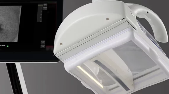
Ivenia ABUS Premium was designed to help streamline the supplemental breast ultrasound workloads and enhance diagnoses by improving ease of use and image reproducibility.
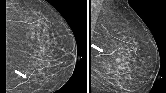
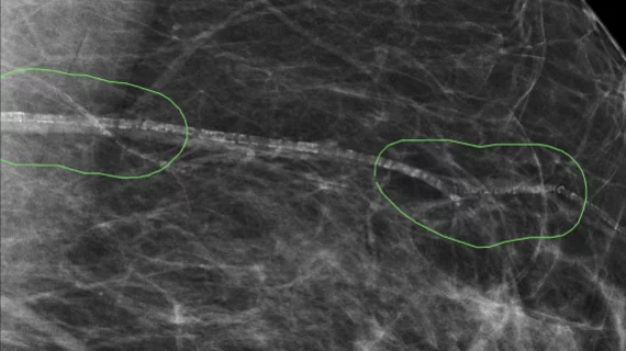
There are no standards requiring radiologists to report on the presence of BACs, even though up to half of referring providers have indicated they would prefer to be made aware of the finding.
Despite the great progress that has been made toward the clinical implementation of AI, new data caution against trusting the technology as a single reader in certain settings.
Jessica Porembka, MD, of the breast imaging division at University of Texas Southwestern Medical Center, said an ultrasound-first strategy for these lesions in DBT is cost-effective and improves efficiency.
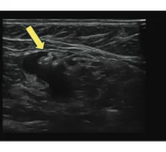
Radiologist Jessica Porembka, MD, FSBI, an associate professor with the breast imaging division at University of Texas Southwestern Medical Center, explains what it means when a mammography report says a patient has architectural distortion.
The postdoctoral research fellow hopes her research will provide greater insight into how the use of oral birth control impacts physical and mental health.
Results from the world’s largest prospective artificial intelligence study revealed the system could significantly benefit breast cancer screening programs.
“POCUS in early pregnancy helps clinicians more efficiently and accurately diagnose problems without compromising the quality of needed first trimester assessments—saving time, money and stress for patients.”
The National Mammography Quality Assurance Advisory Committee provides guidance and recommendations related to the standards by which facilities are regulated.
Clinicians have been using HeartSee to diagnose and treat coronary artery disease since the technology first debuted back in 2018. These latest updates, set to roll out to existing users, are designed to improve diagnostic performance and user access.
The cardiac technologies clinicians use for CVD evaluations have changed significantly in recent years, according to a new analysis of CMS data. While some modalities are on the rise, others are being utilized much less than ever before.