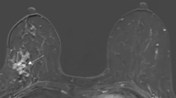
Ultrafast MRI predicts breast cancer upgrade
Preoperative ultrafast MRI could help guide biopsy and surgical management decision in patients with ductal carcinoma in situ (DCIS).
DCIS lesions are frequently upgraded to invasive status following surgery. If radiologists could help to reliably predict whether this will occur prior to surgery, it could aid in surgical planning and spare patients of additional invasive surgeries and procedures such as sentinel lymph node biopsy (SLNB).
Authors of a new paper, published in the American Journal of Roentgenology, suggest that ultrafast MRI (UF-MRI) with conventional dynamic contrast-enhanced (DCE) MRI prior to patients’ breast surgery offers valuable information for planning.
“Preoperative UF-MRI, time to enhancement, and lesion size on conventional dynamic contrast-enhanced (DCE) MRI and mammography show potential in predicting upgrade of ductal carcinoma in situ (DCIS) to invasive cancer at surgery,” first author Rachel Miceli, MD, of NYU Langone Health, and co-authors explained.
The authors analyzed the cases of 68 patients with 68 biopsy-proven pure DCIS lesions. Each of the women had undergone UF-MRI with conventional DCE MRI and had subsequent breast surgery between August 2019 and January 2021. To understand predictors of DCIS upgrade, the experts compared lesion characteristics from mammography, ultrasound, DCE-MRI and UF-MRI to pathological outcomes.
Out of 68 patients, 26 lesions (38%) were upgraded to invasive status from in situ. Upgrade status was significantly associated with shorter time to enhancement (TTE) on preoperative UF-MRI with an optimum threshold of 11 seconds. Larger lesion size (optimum threshold of 4.4 cm) on DCE-MRI and mammography were also associated with upgrade rates.
The sensitivity and specificity of the 11 second TTE threshold was recorded at 76% and 50%. The authors of the study note that these figures bar it from immediate clinical application but do warrant further research.
“The potential for short TTE to predict upgrade of DCIS to invasive cancer is compelling because TTE is clinically available and routinely calculated in clinical practice,” the group wrote, adding that if additional studies with larger cohorts can improve sensitivity and specificity, the metric could be a valuable tool for informing patients and providers on surgical planning.
The study abstract is available here.

