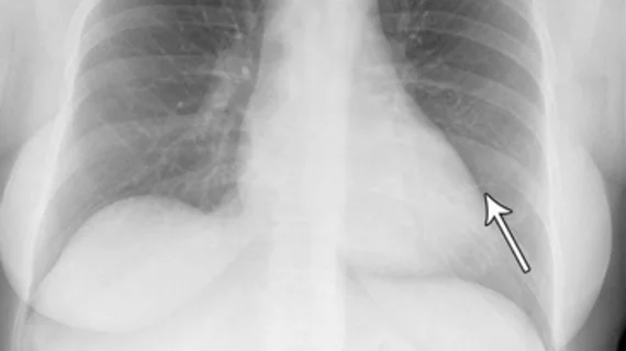
Commercially available AI tool could reduce radiologist workloads by 10% or more
An automated AI tool can differentiate between normal and abnormal chest x-rays, resulting in an impactful workload reduction for radiologists.
That’s according to new research published in Radiology that details an external evaluation of a commercially available AI tool. In a cohort of more than 1,500 patients, the tool achieved a sensitivity of 99.1% for identifying abnormal chest radiographs.
X-rays included in the analysis were performed on patients from emergency departments, in-hospital and outpatient settings. Three thoracic radiologists labeled the radiographs and filed them as either critical, other remarkable, unremarkable or normal (no abnormalities), while the AI tool categorized them as high confidence of normal or abnormal.
The AI tool was able to correctly classify 28% of the images labeled as normal. This equates to approximately 7.8% of the entire cohort that could be safely classified using AI alone.
The tool’s sensitivity was recorded as 99.1% for abnormal radiographs and 99.8% for critical radiographs—better than two board-certified radiologists who also interpreted the exams.
What’s more, the tool was able to identify a wide range of abnormalities, study co-author Louis Lind Plesner, MD, from the Department of Radiology at the Herlev and Gentofte Hospital in Copenhagen, Denmark, shared.
“The most surprising finding was just how sensitive this AI tool was for all kinds of chest disease,” Plesner said. “In fact, we could not find a single chest X-ray in our database where the algorithm made a major mistake. Furthermore, the AI tool had a sensitivity overall better than the clinical board-certified radiologists.”
The tool performed especially well in identifying normal exams in the outpatient setting group. This finding suggests that the tool would be especially beneficial in reducing workloads in such settings, the authors noted.
Given that chest radiographs are one of the most common examinations in both hospitals and outpatient settings, Plesner suggests that there is great potential for validated AI tools, such as the one used for this research (ChestLink, version 2.6; Oxipit), to save radiologists’ time.
“Even a small percentage of automatization can lead to saved time for radiologists, which they can prioritize on more complex matters.”
To learn more, click here.

