Echocardiography

Cardiac ultrasound uses reflected sound waves (echos) to create images of anatomy inside the body. Echocardiograms are the primary cardiac imaging modality used to assess the heart and diagnose or track cardiac issues. Echo is the gold standard imaging modality to assess the heart, particularly with calculating left ventricular ejection fraction (LVEF), which is a measure of cardiac output. In addition to noninvasive standard transthoracic echo (TTE), invasive transesophgeal echo (TEE) is also used when clearer, more detailed imaging of the heart is needed. Both 3D and 4D echo echo systems are rapidly gaining wider adoption and enable new types of assessments, especially in the structural heart space and in transcatheter procedural guidance. Find news on general ultrasound imaging.
Left ventricle-focused echocardiography quick, cost-effective

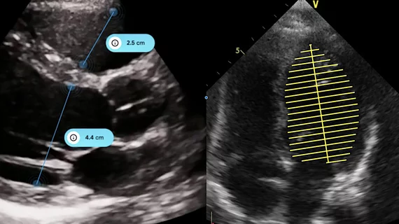
A group of California researchers believes a limited transthoracic echocardiogram (TTE) that solely evaluates the left ventricle could be more efficient and cost-effective in certain patients compared to whole echocardiography.

Thumbnail

Jonathan Lindner elected president of the American Society of Echocardiography

The 17,000-member American Society of Echocardiography (ASE) named Jonathan R. Lindner, MD, president of the organization during its 29th annual scientific sessions in Nashville, Tennessee.

Thumbnail

FDA-approved AI echocardiogram software bests cardiologists in reducing LVEF variability

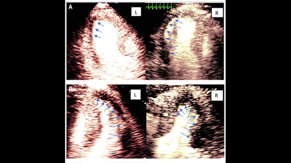
A deep-learning software that can automatically calculate left ventricular ejection fraction (LVEF) with less variability than a cardiologist recently received approval from the U.S. Food and Drug Administration (FDA).

Thumbnail

Is coronary CTA or stress echocardiography better for chest pain triage?

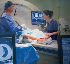
Coronary computed tomography angiography (CTA) is the current go-to for triaging chest pain patients in the emergency department (ED), but a recent study found the often-overlooked stress echocardiography (SE) may actually be safer and provide patients with a better overall experience.

Data fatigue: Machine learning predicts post-echocardiography survival with EHR assistance

Machine learning can accurately predict survival after echocardiography by analyzing unique data produced from heart images and electronic health record (EHR) information, according to a recent study in the Journal of the American College of Cardiology: Cardiovascular Imaging.

New registry seeks to improve cardiovascular ultrasounds

ImageGuideEcho—the first registry devoted solely to measuring quality in cardiovascular ultrasounds— is now open to U.S. physicians.

Thumbnail

Seeing the Big Picture: Training Today’s Imagers to ‘Think Multimodality’

Cardiologists are receiving more exposure to different imaging modalities during their fellowships, but their job prospects and training vary widely. A more comprehensive and multimodality training approach could lead to better results.  

Thumbnail

Genetic variants could be key to identifying chemo-induced cardiotoxicity

As life expectancy continues to expand for cancer patients, clinicians are increasingly dealing with oncological complications like cardiotoxicity, according to a medical team in the Netherlands—and those doctors are met with a paucity of research on the topic.

Around the web

The new technology shows early potential to make a significant impact on imaging workflows and patient care. 

Richard Heller III, MD, RSNA board member and senior VP of policy at Radiology Partners, offers an overview of policies in Congress that are directly impacting imaging.
 

The two companies aim to improve patient access to high-quality MRI scans by combining their artificial intelligence capabilities.