
Low-field MRI systems improve accessibility, but what kind of diagnostic quality do they offer?
MRI scanners with lower magnetic fields promise to increase accessibility to the modality, but there is limited data on whether these scanners are capable of producing diagnostic-quality exams.
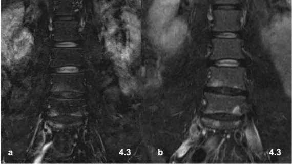
Experts recently compared the use of a 0.55T low-field MRI system to a 1.5T system to determine whether the results rendered were within an acceptable diagnostic range. After reviewing a series of lumbar spine exams completed on healthy patients using both scanners, experts concluded that although the image quality of 0.55T systems is slightly inferior to that produced by 1.5T systems, the low-field scanner can still hold its own in terms of diagnostic value.
The detailed review is published in Academic Radiology, where experts suggest that low-field systems can fill in accessibility gaps known to be barriers to traditional MR imaging.
“Unfortunately, MR imaging remains to be cost-intensive and despite rising numbers of installed scanner systems worldwide, access to this modality is limited,” corresponding author of the new paper Jan Vosshenrich, MD, with the Department of Radiology at University Hospital Basel in Switzerland, and colleagues noted. “A promising opportunity to overcome these limitations is the potential renaissance of low-field MR scanner systems, given advantages in terms of lower costs and fewer demands on infrastructure.”
Low-field systems cost significantly less than high-field scanners and can be implemented in a wider range of settings, which is especially beneficial in resource-poor areas. In the past, the exam quality these systems yield has been subject of criticism, but the experts suggest that improvements in coil development and advances in the use of artificial intelligence have helped to boost the performance of low-field scanners.
In the analysis, healthy volunteers underwent lumbar exams using both systems, with the addition of artificial intelligence-based postprocessing techniques on the 0.55T sequences. With respect to signal/contrast, resolution and assessability of the spinal canal and neuroforamina, three musculoskeletal radiologists rated the 1.5T sequences slightly, but not significantly, higher. Good overall exam quality was reported for both sets of imaging, and good-to-excellent interrater agreement also was achieved.
“Even though image quality was rated slightly lower for all pulse sequences at 0.55T compared to 1.5T MRI, visual assessment by fellowship-trained musculoskeletal radiologists confirmed that lumbar spine imaging can be performed with good to perfect image quality using a low-field MR scanner system,” the group wrote. “Advanced acquisition and post-processing methods at 0.55T allowed to substantially reducing imaging times for the T1-weighted sequence, while only slightly decreasing subjective image quality.”
Although the authors indicate that low-field MRI systems can produce acceptable quality exams of the lumbar spine for the assessment of pain, they suggest that these scanners need to be further tested to determine their feasibility in assessing patients for other specific pathologies, such as nerve root compression and myelopathy.
The study abstract is available here.

In addition to her background in journalism, Hannah also has patient-facing experience in clinical settings, having spent more than 12 years working as a registered rad tech. She began covering the medical imaging industry for Innovate Healthcare in 2021.