Magnetic Resonance Imaging

Magnetic resonance imaging (MRI) is the gold standard imaging modality for soft tissues. It produces detail cross-sectional images of soft tissue and bone anatomy, including muscles, tendons, ligaments, brain and organs, without the use of ionizing radiation. In addition to orthopedic imaging, MRI is also used for heart, brain and breast. MRI uses gadolinium contrast in many exams to highlight tissues and blood vessels, which enhances images and offers better diagnostic quality. It can also be used in conjunction with PET scans. How does MRI work? MR creates images by using powerful magnets to polarize hydrogen atoms in water (the body is made of of more than 80% water) so they face in one direction. A radiofrequency pulse is then used to ping these atoms, causing them to wobble, or resonate. The MRI coils detect this and computers can assemble images from the signals. Basic MRI scans will focus on the resonance of fat and water in two different sequences, which highlight and contrast different features in the anatomy.

Brains are getting bigger—and risk of dementia could be declining

MRI data from the Framingham Heart Study shows an increase in brain volume and surface area over a 40-year timeframe.

Thumbnail

Q&A: MRI accidents are rare, preventable—but safety standardization and certification are key

Emanuel Kanal, MD, founded the American Board of MR Safety to help develop safety standards for MR practices and to certify practitioners.

Thumbnail

Fire at VA hospital in Boston leads to closure of MRI suite

No one was injured in the incident, which originated from under the MRI machine.

SIR 2024: Study finds ultrasound ablation to be a precise, effective treatment for prostate cancer

The MRI-guided transurethral ultrasound ablation comes with minimal side effects, offering cancer patients an alternative to radiation or surgery.

The rapid rise of artificial intelligence (AI) has helped cardiologists, radiologists, nurses and other healthcare providers embrace precision medicine in a way that ensures more heart patients are receiving personalized care.

FDA clears AI-enhanced 3D heart models generated from MR and CT images

The inHeart models significantly reduce cardiac ablation surgery times.

Thumbnail

NIH study finds no evidence of ‘Havana syndrome’ in brains of affected individuals

Brain scans on those with symptoms of the mysterious illness and a control group were found to be the same.

Radiologist using the Philips Smart Quant 3D Neuro artificial intelligence (AI) software to perform our measurements for white matter, gray matter and other parameters on brain MRI.

PHOTO GALLERY of brain imaging

This is a clinical photo gallery of neuro imaging and what conditions can be can be visualized in brain scans, and various imaging techniques used.

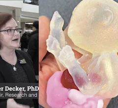
Medical 3D printing expert Summer Decker, PhD, professor and vice chair for research and innovation, and director of 3D clinical applications, University of South Florida (USF) Morsani College of Medicine and Tampa General Hospital, explains what is involved in creating a 3D printiung program in radiology and how it helps patient care.

Pitching the business case for 3D printing labs in radiology

Medical 3D printing expert Summer Decker, PhD, University of South Florida, explains how her lab has helped improve patient care. 

Around the web

The nuclear imaging isotope shortage of molybdenum-99 may be over now that the sidelined reactor is restarting. ASNC's president says PET and new SPECT technologies helped cardiac imaging labs better weather the storm.

CMS has more than doubled the CCTA payment rate from $175 to $357.13. The move, expected to have a significant impact on the utilization of cardiac CT, received immediate praise from imaging specialists.

The newly cleared offering, AutoChamber, was designed with opportunistic screening in mind. It can evaluate many different kinds of CT images, including those originally gathered to screen patients for lung cancer. 

Trimed Popup
Trimed Popup