Magnetic Resonance Imaging

Magnetic resonance imaging (MRI) is the gold standard imaging modality for soft tissues. It produces detail cross-sectional images of soft tissue and bone anatomy, including muscles, tendons, ligaments, brain and organs, without the use of ionizing radiation. In addition to orthopedic imaging, MRI is also used for heart, brain and breast. MRI uses gadolinium contrast in many exams to highlight tissues and blood vessels, which enhances images and offers better diagnostic quality. It can also be used in conjunction with PET scans. How does MRI work? MR creates images by using powerful magnets to polarize hydrogen atoms in water (the body is made of of more than 80% water) so they face in one direction. A radiofrequency pulse is then used to ping these atoms, causing them to wobble, or resonate. The MRI coils detect this and computers can assemble images from the signals. Basic MRI scans will focus on the resonance of fat and water in two different sequences, which highlight and contrast different features in the anatomy.

Thumbnail

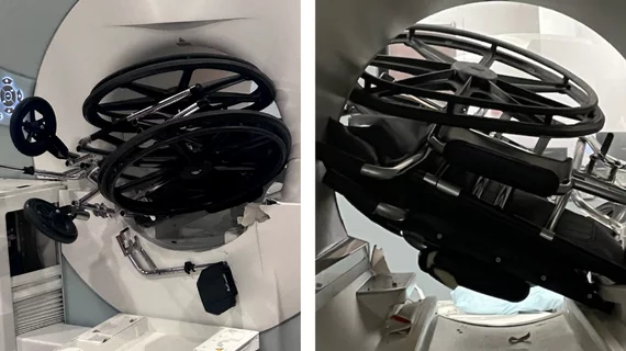
Doctors pilot mobile stroke unit equipped with low-cost MRI machine

Medical University of South Carolina experts believe they are the first to place a portable magnetic resonance imaging unit inside of an ambulance.

New MRI technique earlier detects multiple sclerosis, potentially improving treatment approaches

The tool identified amino acid derivatives that are linked to compromised neuron integrity.

Thumbnail

MRI accurately detects and reveals characteristics of serous borderline ovarian tumors

"Accurate diagnosis and classification of SBOT prior to surgery are crucial for preoperative surgical planning and postoperative treatment," researchers underscored in the European Journal of Radiology.

Thumbnail

MRI should be a standard diagnostic tool for fetuses with known congenital heart disease

The modality can detect certain anomalies difficult to spot using only ultrasound, researchers reported in the Journal of the American College of Cardiology.

Philips MRI

MRI detects 67% of lymph node metastases in patients with prostate cancer, research shows

The modality could be a noninvasive alternative to lymphadenectomy for the detection of cancer spread, experts explained in the European Journal of Radiology.

Thumbnail

Conventional radiography superior to MRI for certain spinal imaging, new research shows

Still, the best results are achieved when the two modalities are combined, according to a study published in Insights Into Imaging.

Thumbnail

Deep learning rivals fellowship-trained radiologists at segmenting breast cancers on MRI

Researchers trained their platforms on more than 60,000 individual breast scans, significantly more than most architectures.

Thumbnail

Machine learning uses MRI to predict lymphovascular invasion in breast cancer patients

These algorithms could fill in where postoperative biopsy sometimes falls short, experts explained in Academic Radiology.

Around the web

GE HealthCare designed the new-look Revolution Vibe CT scanner to help hospitals and health systems embrace CCTA and improve overall efficiency.

Clinicians have been using HeartSee to diagnose and treat coronary artery disease since the technology first debuted back in 2018. These latest updates, set to roll out to existing users, are designed to improve diagnostic performance and user access.

The cardiac technologies clinicians use for CVD evaluations have changed significantly in recent years, according to a new analysis of CMS data. While some modalities are on the rise, others are being utilized much less than ever before.