X-ray

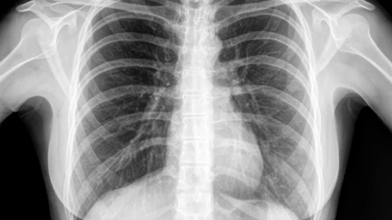
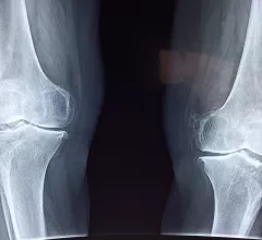
Medical X-rays have mostly converted from film, to computed radiography (CR) that used individual plates to record each X-ray digitally that then had to be uploaded into a PACS, to what is the standard-of-care today with digital radiography (DR). DR X-ray which allows immediate digital transfer of images into a PACS for immediate review. X-rays are used to diagnose fractures, bone abnormalities, lung pathologies and tumors, as well as monitor pediatric growth, plan for surgery and treat oncology patients during radiation therapy. More detailed anatomical imaging, especially soft tissue imaging, is usually sent for advanced imaging with CT or MRI. X-ray, especially mobile DR systems, are a primary use case for artificial intelligence (AI) integration. 

lung cancer pulmonary nodule chest

FDA clears AI-powered software that pinpoints suspicious findings on chest X-rays

VisiRad XR was developed using curated data sets from across the globe, developer Imidex said in an announcement. 

Artificial intelligence (AI) model using chest x-rays to evaluate cardiac function

No echocardiography, no problem? AI evaluates cardiac function using chest X-rays

Researchers think their new AI model could be especially helpful in areas where qualified physicians and/or ultrasound technology are in short supply.

A figure from the study shows a chest radiograph with an area of consolidation involving right lower lung consistent with pneumonia, as well as right pleural effusion. The deep-learning model predicted risk of 30-day mortality of 9%. Right: Gradient- weighted class activation map shows that model prediction was influenced by separate area of image corresponding with heart and liver (yellow and light blue colors). Patient’s CURB-65 score was 4. Patient recovered from pneumonia and remained alive. AJR Image

Deep learning predicts pneumonia mortality on chest X-rays

AI was able to predict 30-day mortality risk predictions more accurately that the current risk assessment.

printable perovskite-based x-ray detector

Printable X-ray detectors are on the horizon

One benefit of these new detectors is that they can operate efficiently across multiple energy levels.

The Nanox ARC cold-cathode, tomosythesis 3D X-ray system is currently pending FDA clearance. If cleared, it would be the first device of its kind to offer cold-cathode X-ray tube technology and the ability to slice through the anatomy in images similar to CT scans aid diagnosis. The new type of tube also could greatly reduce the size and weight of X-ray systems. The vendor sees this system as key to its larger plan to address health disparities and access to imaging world-wide. #Nanox

VIDEO: Cold-cathode X-ray systems may disrupt the medical imaging market

Bhvita Jani, research manager at Signify Research, explains advances in cold-cathode X-ray tube technology and how this might represent a major shift in radiology imaging systems. 
 

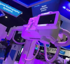
Fixed digital X-ray systems have seen increased market share after a dip in 2020 when hospitals used funding to but more mobile DR systems due to COVID. The Konica-Minolta booth DR system on display at RSNA 2022. Photo by Dave Fornell

VIDEO: Trends in X-ray systems

Bhvita Jani, research manager at Signify Research, explains the technology trends she is seeing in the radiology X-ray market.

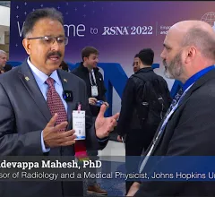
Mahadevappa Mahesh, PhD, professor of radiology and a medical physicist at the Johns Hopkins University School of Medicine, explains a new American College of Radiology (ACR) effort to ensure that lower radiation dose X-ray images under Image Wisely and As Low as Reasonable Achievable (ALARA) meet diagnostic reading standards. He spoke to Radiology Business at the Radiological Society of North America (RSNA) 2022 meeting.

VIDEO: Making sure lower dose X-ray is still diagnostic quality

Mahadevappa Mahesh, PhD, professor of radiology and a medical physicist at the Johns Hopkins University School of Medicine, explains a new ACR effort to ensure that lower radiation dose X-ray images under Image Wisely and As Low as Reasonable Achievable (ALARA) meet diagnostic reading standards.

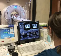
A CT coronary calcium scoring exam at Northwestern Medicine's Central DuPage Hospital in the Chicago Suburbs. Dave Fornell

Trends in the number of radiology studies and a decline in radiation dose

A special report in Radiology offers a rare overview of the number of radiology exams performed each year worldwide and focuses on the decline of associated radiation doses patients receive.

Around the web

RBMA President Peter Moffatt discusses declining reimbursement rates, recruiting challenges and the role of artificial intelligence in transforming the industry.

Deepak Bhatt, MD, director of the Mount Sinai Fuster Heart Hospital and principal investigator of the TRANSFORM trial, explains an emerging technique for cardiac screening: combining coronary CT angiography with artificial intelligence for plaque analysis to create an approach similar to mammography.

A total of 16 cardiology practices from 12 states settled with the DOJ to resolve allegations they overbilled Medicare for imaging agents used to diagnose cardiovascular disease.