X-ray

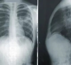
Medical X-rays have mostly converted from film, to computed radiography (CR) that used individual plates to record each X-ray digitally that then had to be uploaded into a PACS, to what is the standard-of-care today with digital radiography (DR). DR X-ray which allows immediate digital transfer of images into a PACS for immediate review. X-rays are used to diagnose fractures, bone abnormalities, lung pathologies and tumors, as well as monitor pediatric growth, plan for surgery and treat oncology patients during radiation therapy. More detailed anatomical imaging, especially soft tissue imaging, is usually sent for advanced imaging with CT or MRI. X-ray, especially mobile DR systems, are a primary use case for artificial intelligence (AI) integration. 

Many clinicians flouting X-ray-first guidelines for ankle imaging

Established clinical guidelines hold that patients presenting with ankle issues should not receive advanced imaging ahead of standard radiography. New research shows a substantial proportion of ordering clinicians sending these patients straight to MRI anyway.

Thumbnail

Deep learning models predict COPD survival based on chest radiographs

Recently, experts explored the efficacy of two deep learning models for grading COPD severity based on chest radiographs—a method that has not yet been thoroughly tested.

Thumbnail

Deep learning model accurately detects COVID-19 on chest X-ray images

Is it pneumonia or COVID-19? With the help of artificial intelligence, a chest X-ray can reveal the answer.

Thumbnail

Explainable AI model accurately auto-labels chest X-rays from open access datasets

A model that can achieve accuracy in line with that of radiologists when labeling open-access datasets could be a key factor to overcoming limitations of artificial intelligence implementation, researchers explained in Nature Communications.

Thumbnail

Intrathoracic complications in COVID patients: Incidence, associations and outcomes

New research published in Clinical Imaging links specific adverse findings on chest imaging of COVID patients to ICU admission, need for intubation and length of hospital stay. 

Thumbnail

Influential pediatric radiologist dies at 89

William Northway Jr., MD, is credited for discovering bronchopulmonary dysplasia on the chest x-rays of premature infants. His research also led to the condition's treatment.

Face masks pulled below the mouth still cause artifacts during dental X-ray exams, experts warn

The authors recommend using metal-free masks, when possible, to avoid the need for repeat imaging.

Thumbnail

Radiologists don’t see eye-to-eye with other providers on fluoroscopic swallow studies

With demand for such exams forecasted to increase alongside an aging population, rads and speech language pathologists do agree that standardization is needed.

Around the web

To fully leverage today's radiology IT systems, standardization is a necessity. Steve Rankin, chief strategy officer for Enlitic, explains how artificial intelligence can help.

RBMA President Peter Moffatt discusses declining reimbursement rates, recruiting challenges and the role of artificial intelligence in transforming the industry.

Deepak Bhatt, MD, director of the Mount Sinai Fuster Heart Hospital and principal investigator of the TRANSFORM trial, explains an emerging technique for cardiac screening: combining coronary CT angiography with artificial intelligence for plaque analysis to create an approach similar to mammography.